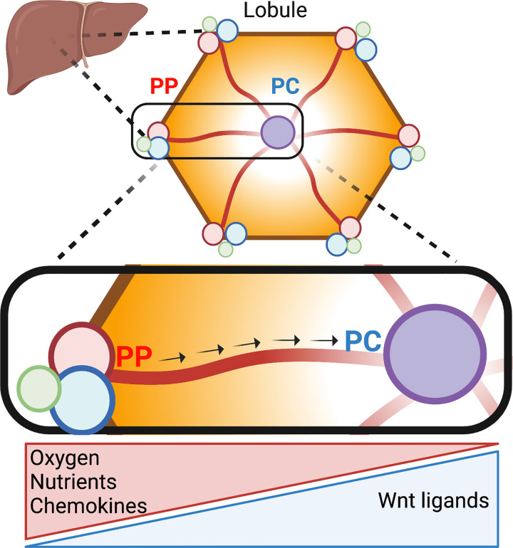
Figure 1.
Liver lobule anatomy and microenvironment. Hepatocytes in the liver are organized in hexagonal units called lobules. The portal triad in the corner of the lobule consists of the portal vein (blue), portal artery (red), and bile duct (green). Blood rich in oxygen and nutrients flows from the periportal toward the pericentral region (arrows). Hepatocytes remove and secrete factors into the bloodstream, thus shaping gradients along the periportal (PP)-pericentral (PC) axis. In addition, nonparenchymal cells, such as immune and endothelial cells, secrete nonuniformly chemokines and Wnt ligands, respectively. As a result, hepatocytes in different parts of the lobule are exposed to distinct microenvironments that determine their gene expression and function. Illustration created using BioRender.

