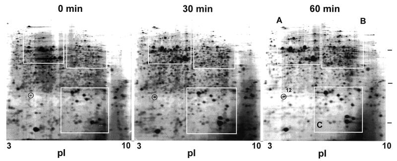
FIG. 1.
2D gel electrophoresis of M. xanthus total proteins before and after heat shock. Spot 12 is circled on the gels. The other heat shock-induced spots in boxes A, B, and C in the 60-min gel are assigned in Fig. 2. Bars at the right are positions of molecular mass markers, 66, 29, and 14 kDa, from the top.

