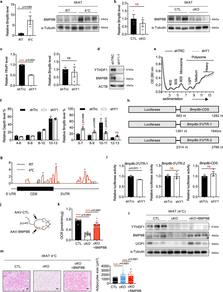
Fig. 5. YTHDF1 regulates BMP8B in an m6A-dependent manner.
a The mRNA and protein levels of BMP8B in iWAT from mice treated at RT or 4 °C. b The mRNA and protein levels of BMP8B in iWAT from Y1CTL and Y1cKO mice treated at 4 °C. c The mRNA level of Ythdf1 and Bmp8b in 3T3-L1 cells with or without YTHDF1 knockdown. d The protein levels of BMP8B expression in 3T3-L1 cells with or without Ythdf1 knockdown. e Polysome profiles of 3T3-L1 cells with or without YTHDF1 knockdown. f The distributions of Gapdh and Bmp8b in polysome fractions. g m6A peak distribution within Bmp8b transcripts. * indicates the predicted m6A peak. h Schematic of luciferase constructs with the predicted m6A sites in the CDS and 3′UTR of Bmp8b mRNA. i Luciferase activity of the Bmp8b reporter in 3T3-L1 cells with or without YTHDF1 knockdown. j Schematic of the mouse treatment regimen. k–m Mice were treated at 4 °C. k Oxygen consumption rates (OCR) of iWAT from Y1CTL and Y1cKO mice with or without BMP8B overexpression. Data were presented as mean ± SEM (n = 6 biologically independent mice). ****P < 0.0001. l The expression of BMP8B and UCP1 in iWAT from Y1CTL and Y1cKO mice with or without BMP8B expression. m H&E staining of iWAT depots from Y1CTL and Y1cKO mice with or without BMP8B expression. Scale bar, 50 μm. The white line indicated the average size. No adjustments. Data in a–c, f, and i were presented as mean ± SEM (n = 3). ns, not significant, *P < 0.05, ****P < 0.0001. All of the P-values are determined by unpaired two-sided t-test. Source data are provided as a Source Data file.

