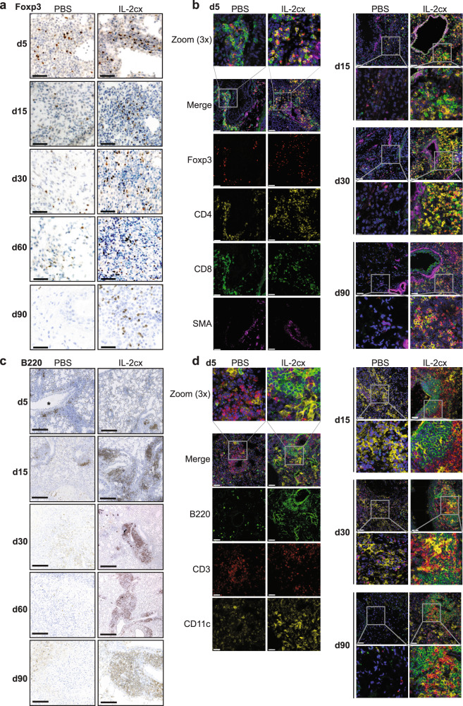
Fig. 5. Localization of immune cell subsets in allografted lungs.
BALB/c lungs were transplanted as in Fig. 1a into C57BL/6 recipients pre-treated as indicated. a Immunohistochemistry staining of Foxp3 in grafted lungs of PBS- (top panels) and IL-2cx-treated mice (bottom panels). Lungs were harvested on indicated days after transplantation. Scale bars = 50 μm. b Immunofluorescence staining for Foxp3 (red), CD4 (yellow), CD8 (green), α-smooth muscle actin (SMA; purple), and nuclei (DAPI; blue) in grafted lungs of PBS- (top panels) and IL-2cx-treated mice (bottom panels). Lungs were harvested on indicated days after transplantation. Scale bars = 50 μm. Squares indicate areas of 3X zoom. c Immunohistochemistry staining for B220 in grafted lungs of PBS- (top panels) and IL-2cx-treated mice (bottom panels). Lungs were harvested on indicated days after transplantation. Scale bars = 300 μm. Magnification ×25. d Immunofluorescence staining for B220 (green), CD3 (red), CD11c (yellow), and nuclei (DAPI; blue) in grafted lungs of PBS- (top panels) and IL-2cx-treated mice (bottom panels). Lungs were harvested on indicated days after transplantation. Scale bars = 50 μm. Squares indicate areas of 3X zoom. Representative images of n ≥ 4 mice of 2–3 independent experiments.

