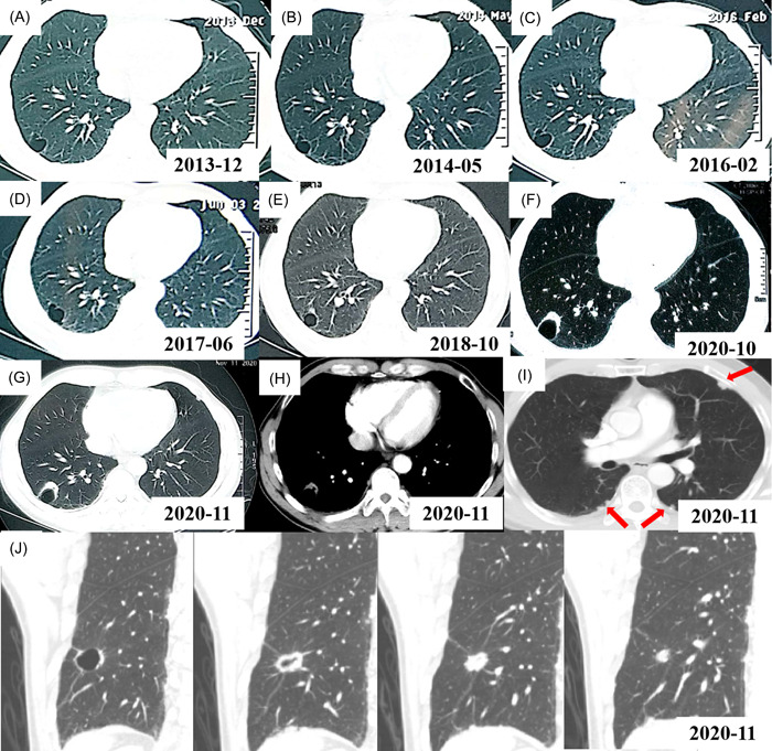
Figure 1.

Computed tomography (CT) scans of the lung cystic airspaces from 2013 to 2020. (A) Initial CT shows a local thin‐walled air cavity in the right lower lobe in Dec 2013. (B–D) On a CT follow‐up, the size of the thin‐walled air cavity changed little in May 2014 (B), Feb 2016 (C), and Jun 2017 (D), respectively. (E) CT shows that a nonsolid nodule appeared adjacent to the wall of cystic airspace in Oct 2018. (F) CT shows that the size of the nodule increased and the wall of the cystic airspace thicken in Oct 2020. (G) CT shows a thicken‐walled cystic space with exophytic solid nodule along the cyst wall in Nov 2020. (H) CT shows a soft tissue nodule in the mediastinal window in Nov 2020. (I) CT shows local pleura irregular thickening and multiple small nodules on both lungs in Nov 2020. (J) CT images of the lung cystic airspace and nodule after three‐dimensional reconstruction in November 2020.
