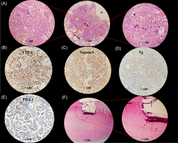
Figure 2.

Histopathology and immumohistochemical staining of main makers. (A) Hematoxylin and eosin (HE) staining of resected lung specimen (*lung cavity; **tumor issue; ***normal lung tissue). (B–E) Tumor cells are positive for TTF‐1 (B) and NapsinA (C), while negative for Tg (D) and PD‐L1 (E, Dako 22C3 antibody). (F) HE staining of the resected pleural nodule.
