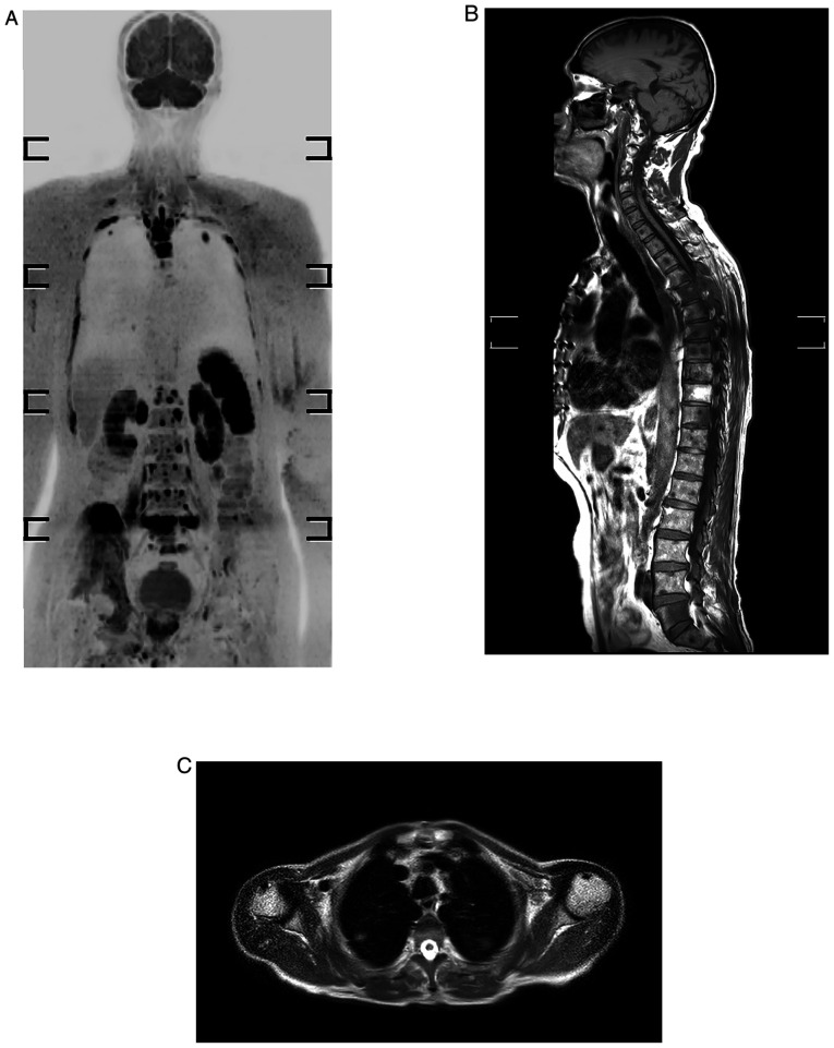
Figure 1.
Whole-body MRI of a 69-year-old male who developed prostate cancer presented with skeletal and lung metastases. (A) Inverted black-white coronal diffusion-weighted MRI with background body signal suppression sequence displaying multiple rib, vertebral and right iliac bone lesions, and also pulmonary lesions at upper lobes bilaterally. (B) T1-weighted image displaying hypointense dorsal and lumbar skeletal lesions. (C) Axial T2-weightedimage showing abnormal focal pulmonary lesions.

