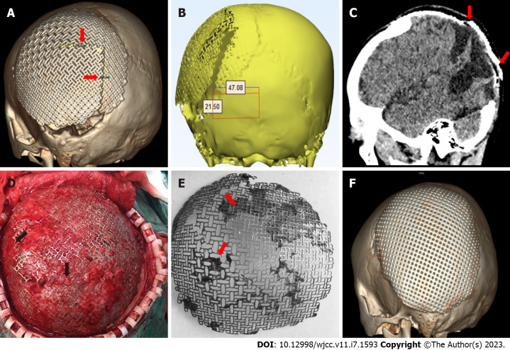
Figure 2.
Images before, during, and after second cranioplasty. A: Three-dimensional (3-D) computerized tomography (CT) reconstruction before the second cranioplasty displayed a reverse “L”-shaped fracture of the titanium mesh prosthesis; B and C: CT at the coronal and sagittal plane revealed the prosthetic fissure (red arrow); D: Intraoperative photograph showing the clear edge of the titanium mesh fracture; E: The fractured titanium mesh was removed during the second cranioplasty; F: 3-D CT reconstruction after the second cranioplasty disclosed an intact and ideally positioned implant.

