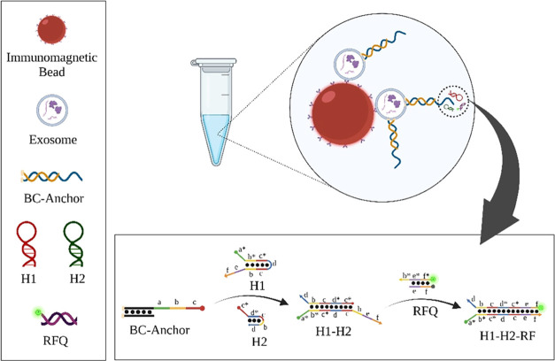
Figure 3.
Schematic illustration of the proposed method for the quantitative evaluation of exosome based on magnetic separation and enzyme-free signal amplification. Anti-CD63 antibodies were immobilized on carboxylic acid-functionalized magnetic beads (MBs). The exosomes sample was mixed with anti-CD63 MBs in a microtube, where the exosomes were captured on the surface of anti-CD63 MBs through antibody–antigen reactions. Subsequently, the BC-anchors were introduced and spontaneously inserted into the lipid bilayer membrane of exosomes captured on anti-CD63 MBs. Region a of the BC-anchor can function as a toehold to trigger interactions with the exposed region a of H1. Next, the newly exposed region c of H1 is free to hybridize with the toehold region c* of H2 to form the H1–H2 duplex. The exposed toehold region of the H1–H2 duplex continues to hybridize with region b* of RFQ. The hybridization triggers the branch migration reaction to displace the RQ quencher, which restores the RF fluorescence signal in the obtained H1–H2–RF complex (adapted from Wang et al.).50

