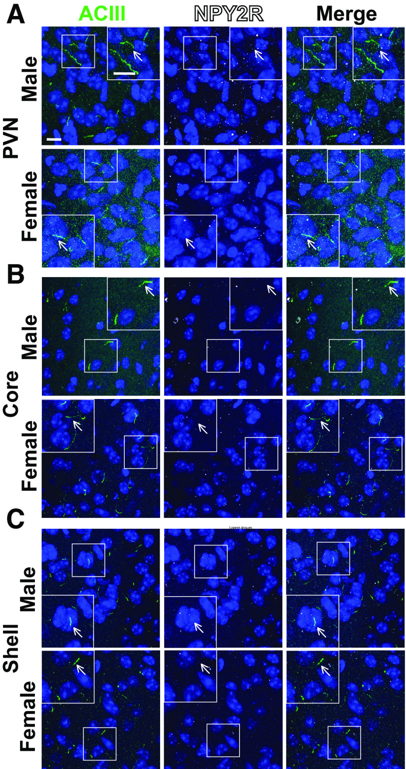
Figure 8.
NPY2R does not localize to cilia in the PVN or nucleus accumbens. A–C, Representative immunofluorescence images of neuronal cilia (ACIII, green) and NPY2R (white) within the PVN (A), nucleus accumbens core (B), and nucleus accumbens shell (C). Scale bars, 10 µm. Hoechst nuclei blue stain was used. Arrows indicate example cilia.

