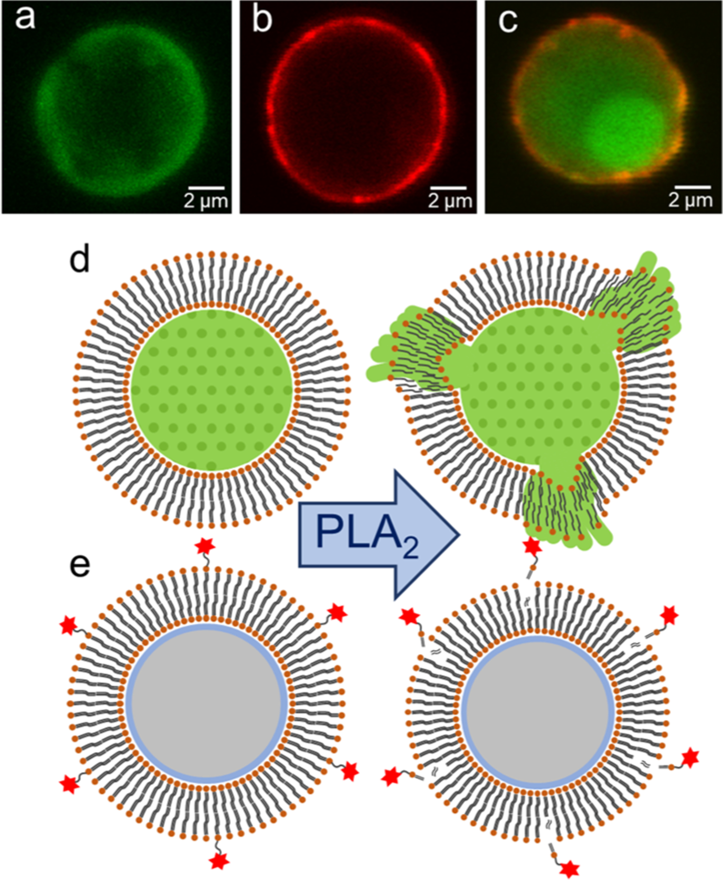
Figure 1.

Formation of fluorescent lipo-beads and the two approaches for detecting membrane–PLA2 interaction. Lipo-beads (a) with a nonfluorescent lipid bilayer with encapsulated fluorescein, (b) with a lipid bilayer comprised of fluorescent lipid, RPE, and (c) with a lipid bilayer comprised of RPE and encapsulated fluorescein. The concept of release of (d) fluorescein dye and (e) fluorescent lipid because of the PLA2-mediated membrane hydrolysis.
