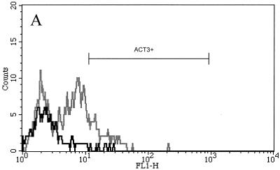
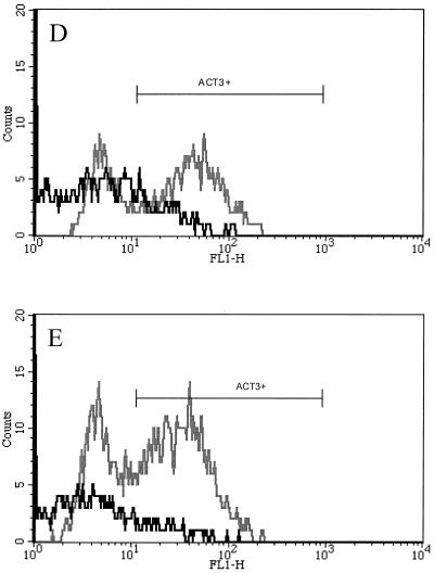
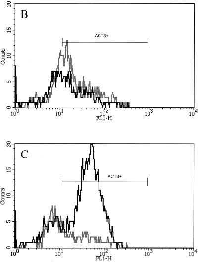
FIG. 1.
Representative flow cytometric profiles of bovine lymphocytes labeled with mouse anti-ACT3 (CACT114A MAb) and anti-CD4 (CACT138A MAb) or anti-CD8 (CACT80C MAb). Bovine PBMC were isolated and stimulated with SEC1 or ConA for 4 or 7 days. In prior experiments, untreated cultures did not express significantly elevated levels of ACT3 (results not shown). (A to D) Cells from untreated control prior to stimulation (A), from 4-day culture with SEC1 (B), from 7-day culture with SEC1 (C), from 4-day culture with ConA (D), and from 7-day culture with ConA (E). The black lines indicate CD8+-T-cell populations, while the gray lines indicate CD4+-T-cell populations colabeled with anti-ACT3 (CACT114A). FL1-H, fluorescence intensity in fluorescence channel 1.


