Figure 3.

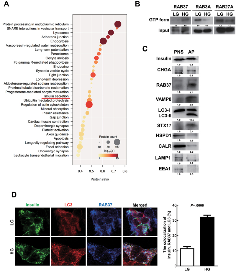
RAB37 was activated and colocalized with insulin and autophagosomes in MIN6 cells under HG treatment. (A) MIN6 cells were treated with HG (25 mM) for 1 h followed by purification of autophagosomes (AP) using density gradient centrifugation. The proteins in the purified autophagosome were identified by HPLC/MS/MS followed by the analysis of Gene Ontology Biological Processes (GOBP). Circular size is proportional to the fold enrichment and the color is proportional to significance. (B) MIN6 cells were treated with LG or HG for 1 h. The levels of active-forms of RAB3A, RAB27A, and RAB37 proteins were determined using extraction beads of active-form RAB proteins or total cellular input followed by immunoblotting. (C) the protein levels of various vesicle markers in the postnuclear supernatant (PNS) and purified autophagosomes (AP) were evaluated by immunoblotting using specific antibodies. (D) Colocalization of insulin (green) with LC3 (red) and RAB37 (blue) in MIN6 cells under LG or HG treatment was investigated under confocal microscopy, N = 120. Scale bars: 10 μm. Student’s t-test was used for statistical analysis.
