
Figure 1.
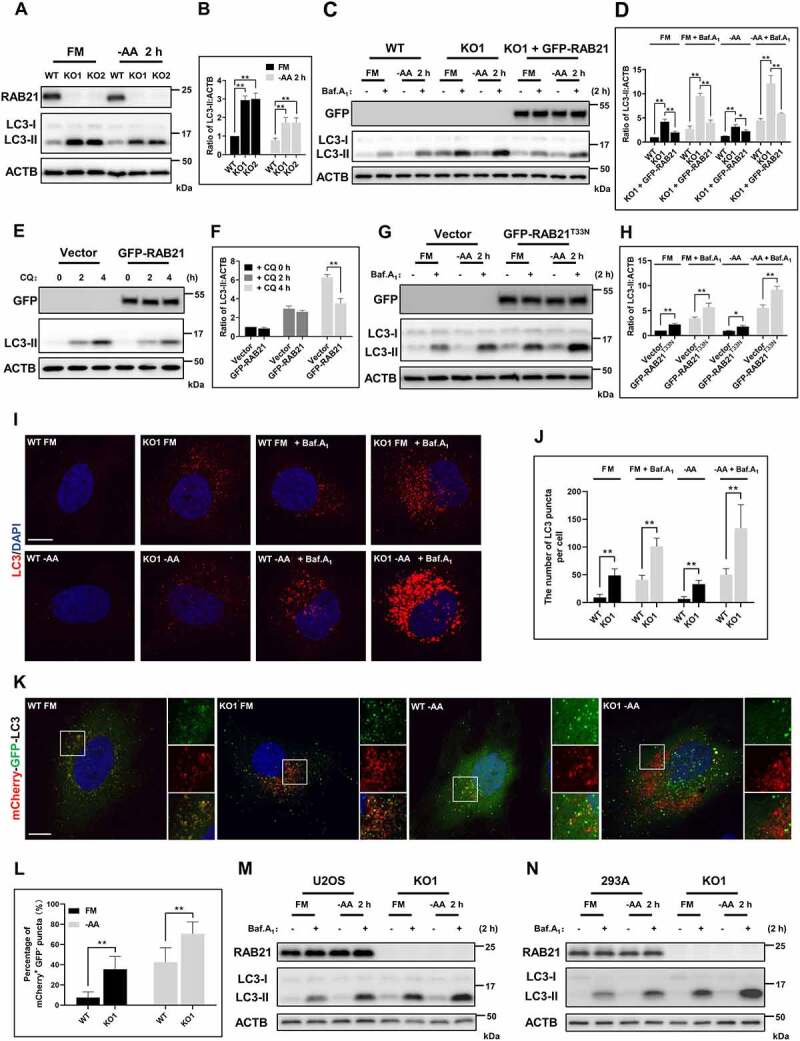
RAB21 regulates basal and starvation-induced autophagic flux. (A-B) WT and RAB21 KO HeLa cells were cultured in full medium (FM) or amino acid-free medium (−AA). The levels of LC3 were examined, and the quantitative results from three independent experiments are shown in B. (C-D) WT, RAB21 KO, and RAB21 KO re-expressing GFP-RAB21 HeLa cells were cultured in FM or -AA with or without 100 nM bafilomycin A1 (Baf.A1). the LC3 levels were examined, and the quantitative results from three independent experiments are shown in D. (E-F) WT and GFP-RAB21-expressing HeLa cells were cultured in FM with or without 50 μM chloroquine (CQ). The LC3 levels were examined, and the quantitative results from three independent experiments are shown in F. (G-H) WT and GFP-RAB21T33N-expressing HeLa cells were cultured in FM or -AA with or without 100 nM Baf.A1. the LC3 levels were examined, and the quantitative results from three independent experiments are shown in H. (I-J) WT and RAB21 KO HeLa cells were cultured in FM or -AA, with or without 100 nM Baf.A1, and LC3 puncta were detected by immunostaining. Scale bar: 10 μm. Quantitative results are shown in J. n > 30 cells from three independent experiments. (K-L) mCherry-GFP-LC3 was expressed in both WT and RAB21 KO HeLa cells cultured in FM or -AA. LC3 puncta were monitored, and the percentage of mCherry+ GFP- puncta (autolysosomes) was quantified in L (n > 35 cells from three independent experiments). Scale bar: 10 μm. (M) WT and RAB21 KO U2OS cells were cultured in FM or -AA with or without 100 nM Baf.A1. (N) WT and RAB21 KO 293A cells were cultured in FM or -AA with or without 100 nM Baf.A1. the graphs express the mean ± SEM. *P <0.05; **P <0.01; NS, not significant.