Figure 4.

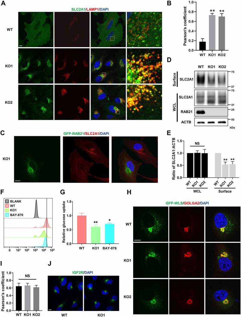
RAB21 KO disrupts retrograde trafficking of SLC2A1 and affects glucose uptake. (A) WT and RAB21 KO HeLa cells were immunostained for SLC2A1 (green) along with LAMP1 (red). (B) Colocalization of SLC2A1 with LAMP1 in a was analyzed by calculation of Pearson’s coefficients (n > 25 cells from three independent experiments). (C) RAB21 KO and RAB21 KO re-expressing GFP-RAB21 HeLa cells were mixed and seeded onto coverslips for SLC2A1 (red) immunostaining. (D-E) WT and RAB21 KO HeLa cells were surface-biotinylated, followed by streptavidin isolation and immunoblot analysis. Surface, cell surface proteins. WCL, whole cell lysate. The quantitative results from three independent experiments are shown in E. (F-G) WT and RAB21 KO HeLa cells were cultured with 100 μM 2-NBDG for 4 h, and 2-NBDG uptake was measured by flow cytometry. WT HeLa cells pretreated with 10 μM BAY-876 for 24 h were used as a positive control. The results from three independent experiments were statistically analyzed in G. (H-I) GFP-WLS was expressed in WT and RAB21 KO HeLa cells. The colocalization of GFP-WLS with GOLGA2 was detected by immunostaining. The Pearson’s coefficients (n > 20 cells from three independent experiments) are shown in I. (J) the subcellular localizations of endogenous IGF2R in WT and RAB21 KO HeLa cells were detected by immunostaining. Scale bars: 10 μm. The graphs express the mean ± SEM. *P <0.05; **P <0.01; NS, not significant.
