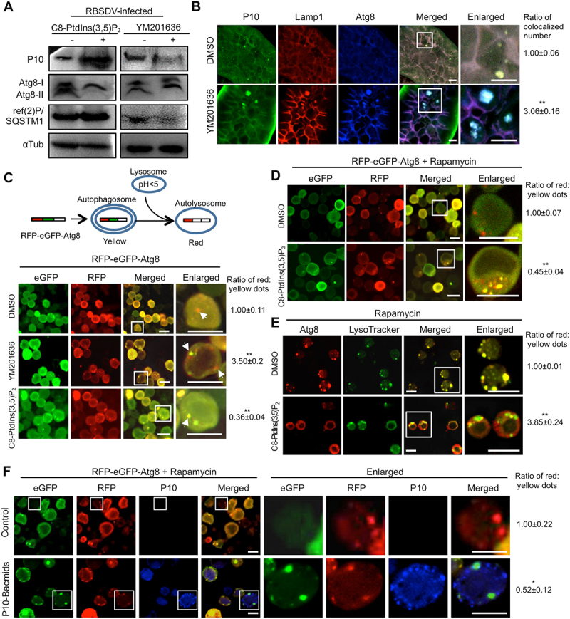
Figure 5.

PtdIns(3,5)P2 inhibits autophagy by blocking lysosome-autophagosome fusion. (A) PtdIns(3,5)P2 regulated the autophagy level in L. striatellus. After acquiring the virus for 2 days, the nymphs of L. striatellus were injected with DMSO, YM201636 (20 μM), or C8-PtdIns(3,5)P2 (0.5 μM). The relative abundances of P10, Atg8, and ref(2)P/SQSTM1 in L. striatellus were detected by western blotting at 10 d post microinjection. (B) YM201636 treatment caused the colocalization of RBSDV, lysosomes, and autophagosomes. After acquiring the virus for 6 days, the nymphs of L. striatellus were injected with DMSO or YM201636 (20 μM) and collected at 2 d post microinjection. The colocalization of RBSDV, lysosome, and autophagosome in the intestines of L. striatellus was analyzed by immunofluorescence analysis with conjugated antibodies RBSDV-FITC, Lamp1-rhodamine, and Atg8-Alexa Fluor 647. (C) PtdIns(3,5)P2 regulated the lysosome-autophagosome fusion in Sf9 cells. Sf9 cells were infected with RFP-eGFP-Atg8 bacmids for 6 h, and then incubated with DMSO, YM201636 (100 nM), or C8-PtdIns(3,5)P2 (50 nM) for 6 h. The cells were viewed by confocal microscopy at 36 h post reagents treatment. The white arrows indicated the red and yellow dots in the cells. (D-E) PtdIns(3,5)P2 inhibited rapamycin-induced lysosome-autophagosome fusion in Sf9 cells. The Sf9 cells were transfected with RFP-eGFP-Atg8 bacmids for 6 h and treated with rapamycin (20 μM) for another 6 h. The cells were rinsed with PBS buffer and then incubated with DMSO or C8-PtdIns(3,5)P2 (50 nM) for 12 h. The expression of RFP-eGFP-Atg8 in the Sf9 cells was analyzed by a confocal microscopy (D). Sf9 cells were treated with rapamycin for 6 h, and then incubated with DMSO or C8-PtdIns(3,5)P2 (50 nM) for 6 h. The Atg8 and lysosomes in Sf9 cells were labeled by immunofluorescence analysis with Atg8-rhodamine antibody and LysoTracker (E). (F) P10 protein blocks the fusion of autophagosome and lysosome. The Sf9 cells were infected with RFP-eGFP-Atg8 bacmids and P10 bacmids together for 6 h, and then treated with 20 μM rapamycin for 6 h. After 24 h culture in complete medium, the expression of GFP, RFP, and P10 was detected by immunofluorescence analysis. RBSDV monoclonal antibody conjugated Alexa Fluor 647 was used to detect P10 protein. The numbers of yellow dots and red dots were counted in 150 cells with distinct cell contours and containing punctate fluorescent dots in each treatment, and then the numbers of red:yellow dots was analyzed. The ratio of red:yellow dot numbers in the cells treated with YM201636 or C8-PtdIns(3,5)P2 was normalized to that in the DMSO control. Scale bars, 10 μm. Error bars, mean ± SE. The quantification was analyzed by Student’s t-test. *, p <0.05; **, p <0.01.
