. 2023 Jan 30;31(1):19–28. doi: 10.5606/tgkdc.dergisi.2023.23396

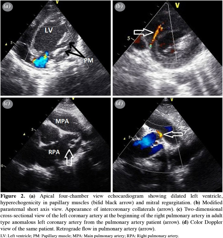
Figure 2. (a) Apical four-chamber view echocardiogram showing dilated left ventricle, hyperechogenicity in papillary muscles (bifid black arrow) and mitral regurgitation. (b) Modified parasternal short axis view. Appearance of intercoronary collaterals (arrow). (c) Two-dimensional cross-sectional view of the left coronary artery at the beginning of the right pulmonary artery in adult type anomalous left coronary artery from the pulmonary artery patient (arrow). (d) Color Doppler view of the same patient. Retrograde flow in pulmonary artery (arrow). LV: Left ventricle; PM: Papillary muscle; MPA: Main pulmonary artery; RPA: Right pulmonary artery.

Figure 2