. 2023 Jan 30;31(1):19–28. doi: 10.5606/tgkdc.dergisi.2023.23396

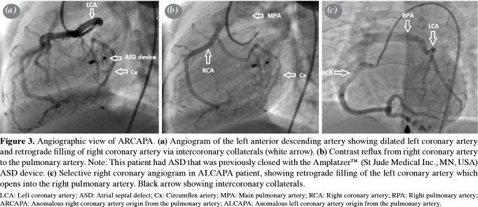
Figure 3. Angiographic view of ARCAPA. (a) Angiogram of the left anterior descending artery showing dilated left coronary artery and retrograde filling of right coronary artery via intercoronary collaterals (white arrow). (b) Contrast reflux from right coronary artery to the pulmonary artery. Note: This patient had ASD that was previously closed with the AmplatzerTM (St Jude Medical Inc., MN, USA) ASD device. (c) Selective right coronary angiogram in ALCAPA patient, showing retrograde filling of the left coronary artery which opens into the right pulmonary artery. Black arrow showing intercoronary collaterals. LCA: Left coronary artery; ASD: Atrial septal defect; Cx: Circumflex artery; MPA: Main pulmonary artery; RCA: Right coronary artery; RPA: Right pulmonary artery; ARCAPA: Anomalous right coronary artery origin from the pulmonary artery; ALCAPA: Anomalous left coronary artery origin from the pulmonary artery.LV: Left ventricle; PM: Papillary muscle; MPA: Main pulmonary artery; RPA: Right pulmonary artery.

Figure 3