Abstract
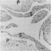
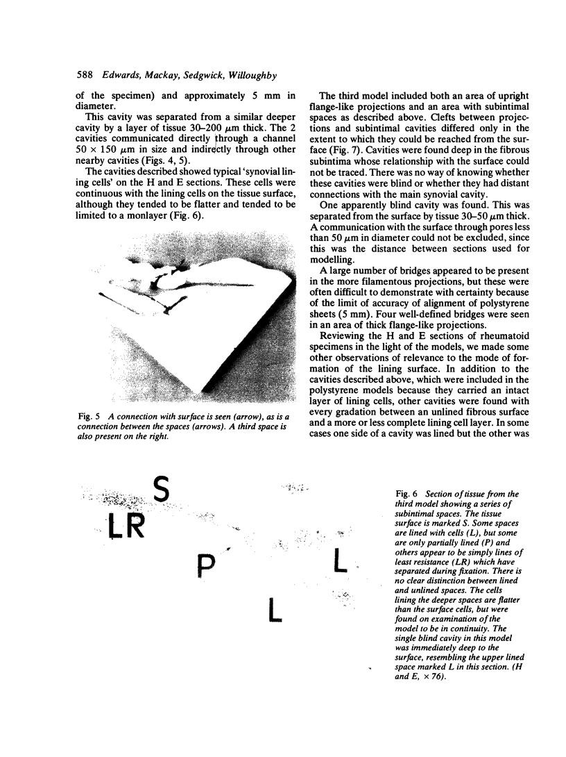
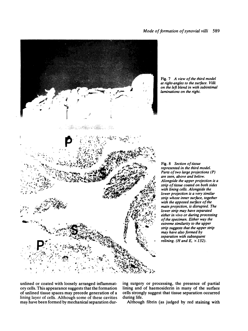
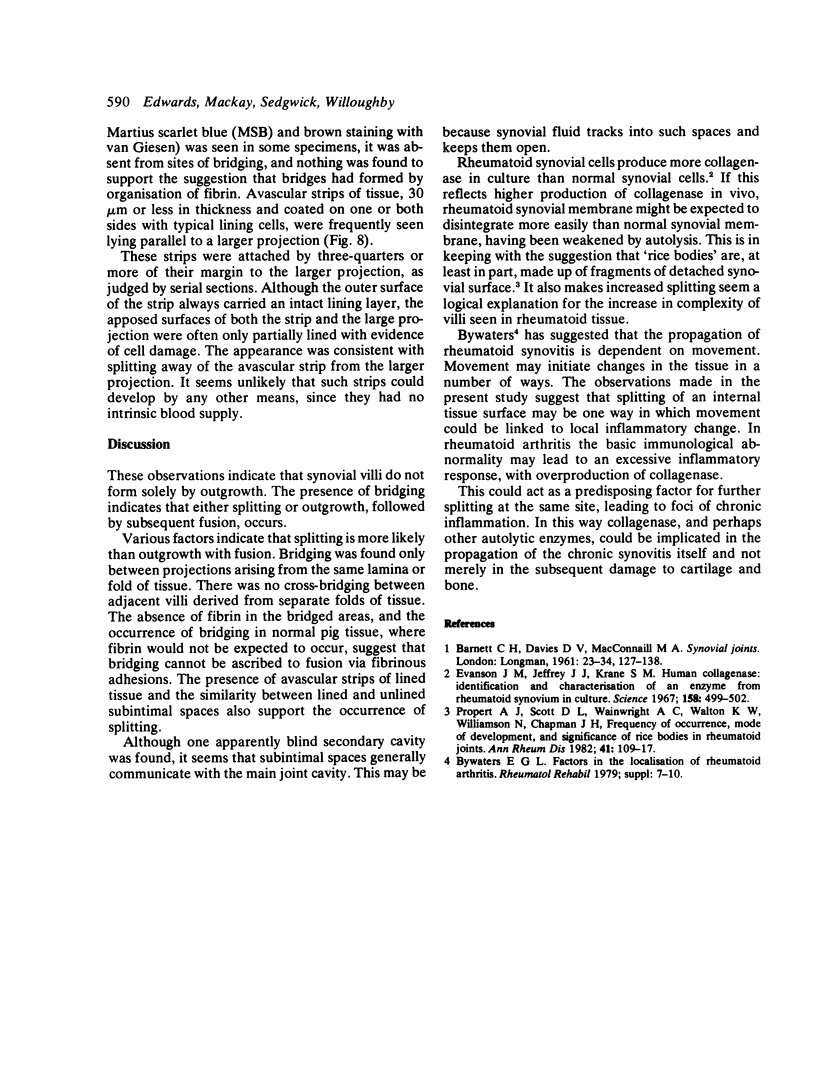
Synovial tissue from 6 normal pigs and from 16 patients undergoing arthrotomy for joint disease was examined by dissecting microscopy. Scale models were constructed of 3 human synovial specimens from photographic magnifications of serial sections. Surface bridging and subintimal cavitation were observed, particularly in tissue from patients with rheumatoid arthritis. These features suggest that synovial surface projections (villi) do not form simply by outgrowth. Reference to original haematoxylin and eosin stained sections suggested that tissue splitting contributes to the formation of villi.
Full text
PDF
Images in this article
Selected References
These references are in PubMed. This may not be the complete list of references from this article.
- Bywaters E. G. Factors in the localization of rheumatoid arthritis. Rheumatol Rehabil. 1979;Suppl:7–10. doi: 10.1093/rheumatology/xviii.suppl.7. [DOI] [PubMed] [Google Scholar]
- Evanson J. M., Jeffrey J. J., Krane S. M. Human collagenase: identification and characterization of an enzyme from rheumatoid synovium in culture. Science. 1967 Oct 27;158(3800):499–502. doi: 10.1126/science.158.3800.499. [DOI] [PubMed] [Google Scholar]
- Popert A. J., Scott D. L., Wainwright A. C., Walton K. W., Williamson N., Chapman J. H. Frequency of occurrence, mode of development, and significance or rice bodies in rheumatoid joints. Ann Rheum Dis. 1982 Apr;41(2):109–117. doi: 10.1136/ard.41.2.109. [DOI] [PMC free article] [PubMed] [Google Scholar]


