Abstract
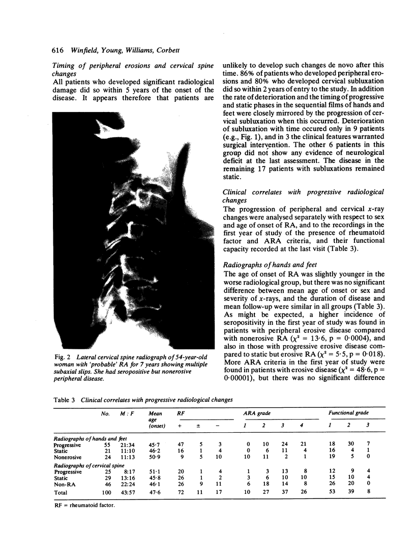
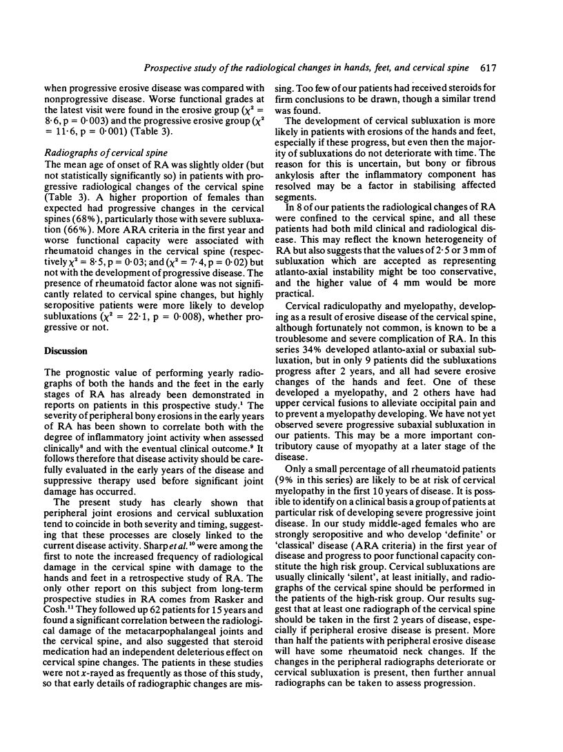
Annual radiographs of hands, feet, and cervical spine were taken in 100 patients with rheumatoid arthritis from the first year of disease for a mean follow-up period of 9.5 years. Seventy-six patients developed peripheral erosive disease and 54 developed rheumatoid changes of the cervical spine, of whom 34 (63%) had subluxations. The severity of rheumatoid neck damage correlated strongly with the severity of peripheral erosive disease (p = 0.002). Cervical subluxation was more likely to occur in patients with erosions of the hands and feet which deteriorated progressively with time (p = 0.018). The timing and severity of cervical subluxation coincided with the progression of peripheral erosive disease in 26 of these 34 patients (76.5%). The other 8 patients with cervical subluxation (23.5%) had none or only mild peripheral erosions, but their subluxations did not progress with time. There were 9 patients with marked cervical subluxations which deteriorated relentlessly, and they all also had severe progressive erosive disease of the hands and feet. One of these patients developed a cervical myelopathy, and 2 other patients with normal neurological signs had upper cervical fusions performed for severe occipital headache. This small group of rheumatoid patients who are at risk of developing cervical myelopathy cannot be predicted with certainty, but can be selected out at an early stage by performing regular radiographs of hands, feet, and cervical spine.
Full text
PDF
Images in this article
Selected References
These references are in PubMed. This may not be the complete list of references from this article.
- Brook A., Corbett M. Radiographic changes in early rheumatoid disease. Ann Rheum Dis. 1977 Feb;36(1):71–73. doi: 10.1136/ard.36.1.71. [DOI] [PMC free article] [PubMed] [Google Scholar]
- Brook A., Fleming A., Corbett M. Relationship of radiological change to clinical outcome in rheumatoid arthritis. Ann Rheum Dis. 1977 Jun;36(3):274–275. doi: 10.1136/ard.36.3.274. [DOI] [PMC free article] [PubMed] [Google Scholar]
- DIAGNOSTIC criteria for rheumatoid arthritis: 1958 revision by a committee of the American Rheumatism Association. Ann Rheum Dis. 1959 Mar;18(1):49–53. [PMC free article] [PubMed] [Google Scholar]
- Fleming A., Crown J. M., Corbett M. Early rheumatoid disease. I. Onset. Ann Rheum Dis. 1976 Aug;35(4):357–360. doi: 10.1136/ard.35.4.357. [DOI] [PMC free article] [PubMed] [Google Scholar]
- Rasker J. J., Cosh J. A. Radiological study of cervical spine and hand in patients with rheumatoid arthritis of 15 years' duration: an assessment of the effects of corticosteroid treatment. Ann Rheum Dis. 1978 Dec;37(6):529–535. doi: 10.1136/ard.37.6.529. [DOI] [PMC free article] [PubMed] [Google Scholar]
- SHARP J., PURSER D. W., LAWRENCE J. S. Rheumatoid arthritis of the cervical spine in the adult. Ann Rheum Dis. 1958 Sep;17(3):303–313. doi: 10.1136/ard.17.3.303. [DOI] [PMC free article] [PubMed] [Google Scholar]
- Winfield J., Cooke D., Brook A. S., Corbett M. A prospective study of the radiological changes in the cervical spine in early rheumatoid disease. Ann Rheum Dis. 1981 Apr;40(2):109–114. doi: 10.1136/ard.40.2.109. [DOI] [PMC free article] [PubMed] [Google Scholar]
- Young A., Corbett M., Brook A. The clinical assessment of joint inflammatory activity in rheumatoid arthritis related to radiological progression. Rheumatol Rehabil. 1980 Feb;19(1):14–19. doi: 10.1093/rheumatology/19.1.14. [DOI] [PubMed] [Google Scholar]


