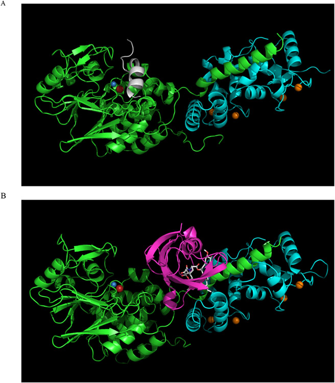
Figure 8. Inhibition of PP2B by immunophilin/immunosuppressant complex.
Inhibition of PP2B by immunophilin/immunosuppressant complex. A. The structure of a PP2B holoenzyme is shown in a cartoon representation. The PP2B catalytic subunit is green, the regulatory B subunit is cyan, and the autoinhibitory domain is white. Metal ions are shown as spheres: Fe is red, Zn is blue, and Ca is orange. B. The structure of a PP2B holoenzyme bound to the inhibitory FKBP12:tacrolimus complex is shown in a cartoon representation. The PP2B catalytic subunit is green, the regulatory B subunit is cyan, and FKBP12 is magenta. Metal ions are shown as spheres: Fe is red, Zn is blue, and Ca is orange. Tacrolimus (FK506) is shown as sticks

