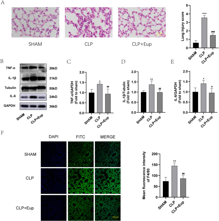
Figure 5.
Effects of Eupatilin on the expression of inflammation indicators and macrophage infiltration in lung tissue of septic mice. (A) Following H&E staining, pictures of the lung tissue in each group were taken using an optical microscope (400 ×, scale bar: 25 µm). Lung injury was scored by observing the degree and extent of lung injury in the sections. (B-E) The protein expression of TNF-α, IL-1β, IL-6 in the lung tissue were analyzed by Western blot. (F) The fluorescence images of F4/80 (green) in lung tissue (scale bar: 100 µm) were captured under the confocal laser microscope. n≥3 per group, one-way ANOVA test. *p < 0.05, **p < 0.01, ***p < 0.001, versus SHAM group; #p < 0.05, ##p < 0.01, ###p < 0.001, versus CLP group.

