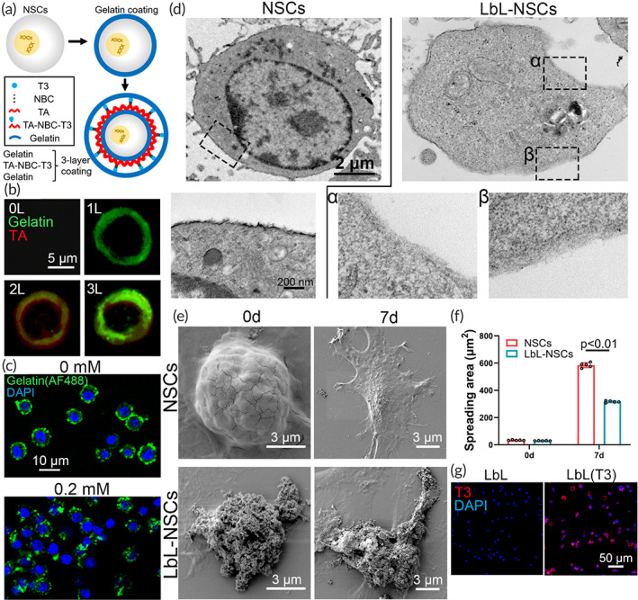
FIGURE 1.

Characterization of layer‐by‐layer (LbL) nanogel. (a) Scheme of the process of LbL nanoencapsulation. (b) Neural stem cells (NSCs) without coating, NSCs coated with AF488‐labeled gelatin, AF488‐labeled gelatin/TA, and AF488‐labeled gelatin/TA/AF488‐labeled gelatin were observed with a confocal microscope. (c) LbL nanogels constructed with AF488‐labeled gelatin were treated with 0 or 0.2 mM H2O2 and examined with a confocal microscope. (d) NSCs and LbL nanogels (indicated as “LbL‐NSCs”) were examined by transmission electron microscopy (TEM). The images in the lower row are magnified views of the inserts in the upper row. (e) NSCs and nanogels seeded on cover slides were observed by scanning electron microscopy (SEM) on Days 0 and 7. The spreading area was calculated in (f) (n = 5). (g) Nanogel and T3‐incorporated nanogel were stained with anti‐T3.
