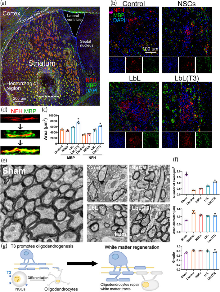
FIGURE 6.

White matter injury and regeneration following neural stem cell (NSC) transplantation in intracerebral hemorrhage (ICH). (a) Immunostaining of brain slices from ICH mice. Perihematomal sites surrounding the framed area were further demonstrated in (b) for white matter regeneration. (b) On Day 21, brain sections of mice from the control, NSC, LbL, and LbL(T3) groups underwent fluorescence staining with anti‐MBP and anti‐NFH. Split channels were placed under each image. (c) MBP‐positive regions and NFH‐positive regions were quantified. *p < 0.01 when LbL(T3) versus control, NSC or LbL groups on Day 21 (n = 6). (d) Regeneration process of the white matter tract. (e) TEM images of perihematomal tissues in ICH from the control, NSC, LbL, and LbL(T3) groups on Day 21. Analysis of the axon number, diameter and G‐ratio is shown in (f). *p < 0.05 when LbL (T3) versus control, NSC or LbL groups (n = 6). (g) Scheme of oligodendrogenesis directed by released T3 and white matter regeneration by oligodendrocytes
