Fig. 3.
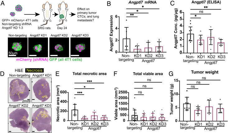
Suppression of Angptl7 normalizes tumor necrosis. (A) Experimental schema. 4T1 tumor cells labeled with membrane GFP and transduced with Angplt7 shRNA or nontargeting control were orthotopically transplanted into a single mammary fat pad of SRG rats. shRNA contained an mCherry tag, so cells expressing shRNA have cytoplasmic mCherry label. Blood, tumor, and lungs were collected. Nontargeting control (n = 11), Angptl7 knockdown KD1 (n = 7), Angptl7 KD2 (n = 6), Angptl7 KD3 (n = 7). (B) In vivo knockdown confirmation by qPCR based on tumor core. (C) ELISA confirmation by in vivo knockdown. Lysates were made from tumor necrotic core from Angptl7 knockdown and nontargeting control tumor cell transplantation experiments. (D) Representative hematoxylin and eosin (H&E) staining of primary tumors for Angptl7 knock down tumors and nontargeting control. Yellow borders indicate the necrotic regions. (E and F) Total necrotic area and total viable area based on H&E staining of Angptl7 KD and nontargeting control tumor. (G) Tumor weight of Angptl7 KD and nontargeting control tumor. All graphs shown display mean ± SD. Mean values shown on graphs. P-values for B and C determined by Mann–Whitney test; for E–G determined by ANOVA. *P <0.05, **P < 0.01, ***P < 0.001, and ****P < 0.0001.

