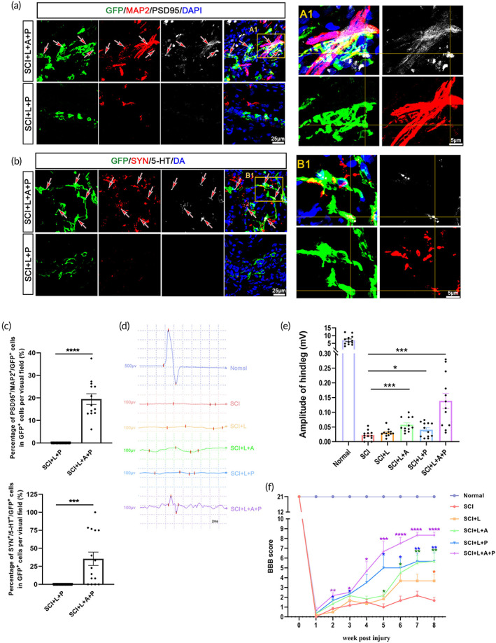
FIGURE 6.

The hscNT structurally integrates with the host neural circuits and effectively improves hindlimb motor function. (a) Host dendrites (GFP−/MAP2+) regenerate into the lesion site and form appositional contacts (arrows) and synapses (PSD95) with the implanted donor neurons (GFP+). (A1) Enlarged image of synaptic junctions between host dendrites and the hscNT. The areas in the yellow boxes show host dendrites. (b) 5‐HT‐labeled host serotonergic axons enter the lesion center and establish a synaptic junction (SYN) with the donor neuron (labeled with GFP). (B1) Enlarged image showing 5‐HT+ axons regenerated in the lesion site and contacting the hscNT. (c) Quantification of synaptic connections between host cells and transplanted cells (GFP+/MAP2+/PSD95+, n = 13 images; GFP+/5‐TH+/SYN+, n = 15 images). (d) CMEP responses in the different groups. (e) Histogram showing that rats implanted with the hscNT have greater CMEP amplitudes (n = 12 images). (f) BBB motor scores after hscNT transplantation in rats with SCI (n = 6 rats). Error bars represent the standard error. Two‐group comparisons were analyzed using Student's t‐test, and multiple groups comparisons were analyzed using one‐way analysis of variance with Tukey's test. NS indicates no significant difference, *p < 0.05; ***p < 0.001; ****p < 0.0001.
