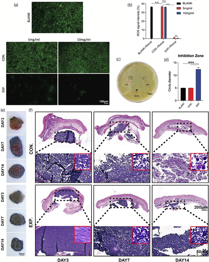
FIGURE 4.

Evaluation of ROS‐scavenging, antibacterial effect, and degradation of granular gel. (a) Fluorescence images and the statistical data (b) of different concentrations of HA MGs and HA‐LA granular gel co‐cultured with HUVECs cells, with the addition of reactive oxygen stimulant (Rosup) (scale bar: 100 μm, n = 3). (c) Antibacterial test of the three groups co‐cultured with Staphylococcus aureus (S aureus) (scale bar: 5 mm, n = 3). (d) Quantification of bacterial inhibition diameter in the three groups co‐cultured with S aureus; General observation (e) and H&E staining (f) of HA MGs and HA‐LA granular gel on days 3, 7, and 14 (CON. = treated with HA MGs; EXP. = treated with HA‐LA granular gel) (scale bar = 200 μm, 50 μm, n = 3). The data represent the mean ± SD. ***P < .001, according to t‐test. HA‐LA, hyaluronic acid‐g‐lipoic acid; HA MGs, hyaluronic acid microgels; HUVECs, Human umbilical vein endothelial cells; SD, standard deviation
