
FIGURE 6.
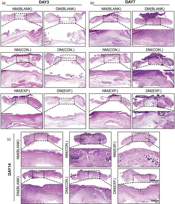
Histology evaluation (H&E staining) of wound healing. (a) H&E staining of the NM (BLANK), NM (CON.), NM (EXP.), DM (BLANK), DM (CON.), and DM (EXP.) groups on day 3, (b) day 7, and (c) day14 (NM (CON.) = normal mice wound treated with HA MGs, NM (EXP.) = normal mice wound treated with HA‐LA granular gel, DM (CON.) = diabetic mice wound treated with HA MGs, DM (EXP.) = diabetic mice wound treated with HA‐LA granular gel) (scale bar: 100 μm and 200 μm, n = 3). HA‐LA, hyaluronic acid‐g‐lipoic acid; HA MGs, hyaluronic acid microgels