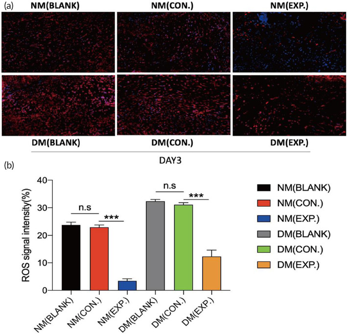
FIGURE 8.

In vivo evaluation of ROS‐scavenging ability of HA MGs and HA‐LA granular gel. (a) Fluorescence images and the statistical data (b) of dihydroethidium (DHE) from different groups on day 3; DAPI (blue) staining of nuclei (NM (CON.) = normal mice wound treated with HA MGs, NM (EXP.) = normal mice wound treated with HA‐LA granular gel, DM (CON.) = diabetic mice wound treated with HA MGs, DM (EXP.) = diabetic mice wound treated with HA‐LA granular gel) (scale bar: 50 μm, n = 3). The data represent the mean ± SD. ***P < .001, according to t‐test. HA‐LA, hyaluronic acid‐g‐lipoic acid; HA MGs, hyaluronic acid microgels; SD, standard deviation
