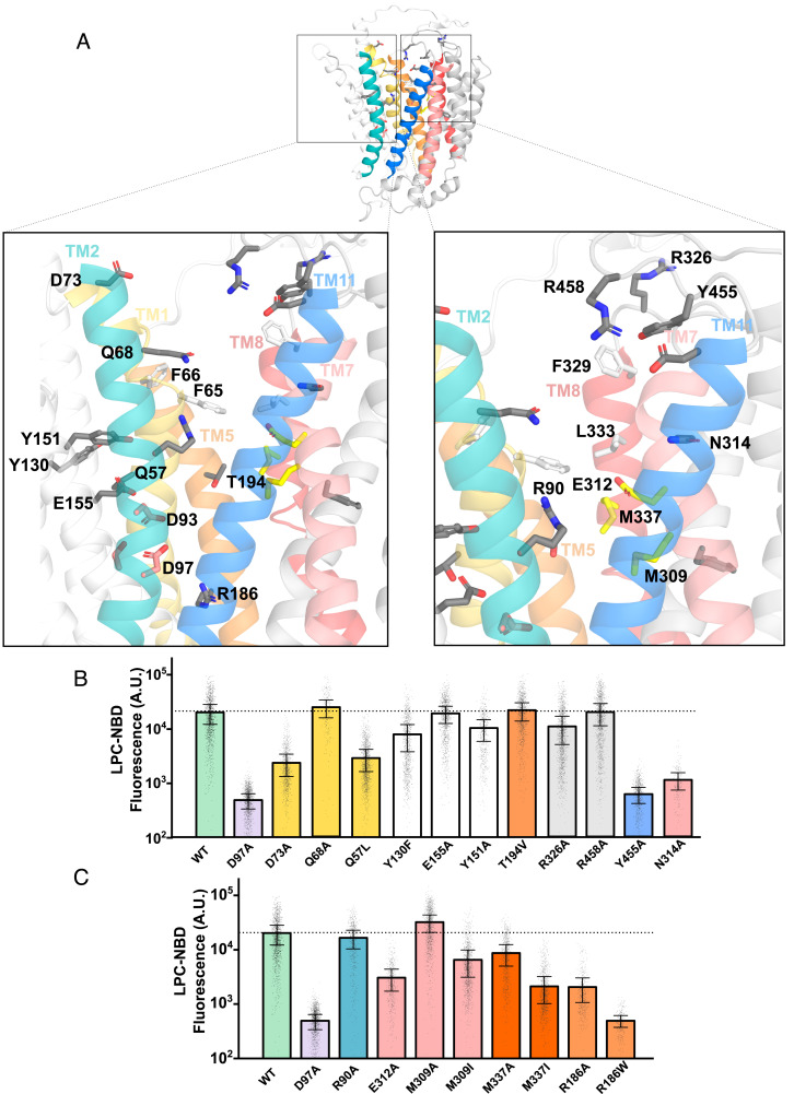
Fig. 5.
Testing the importance of hydrophilic residues lining the central core of Mfsd2a for LPC transport. (A) The model of the outward-open gMfsd2a structure (PDB: 798N). TM2 is in teal and TM11 in blue, TM5 in orange and TM8 in pink, and TM1 in yellow and TM7 in light pink. Residues mutated are shown in gray sticks; hydrophobic residues previously shown to be important for transport (F65, F66, F329, and L333) are shown in white sticks; residues E312, M309, and M337 are shown in yellow sticks. Residues are labeled with human Mfsd2a numbering. (B and C) Quantification of transport of indicated mutants using a single-cell Flow cytometry assay. Data are represented as the mean ± SD, with each dot representing a single analyzed cell with approximately 1,000 cells analyzed per construct. The dotted line represents the average transport level of WT. See SI Appendix, Fig. S4 for primary flow data.

