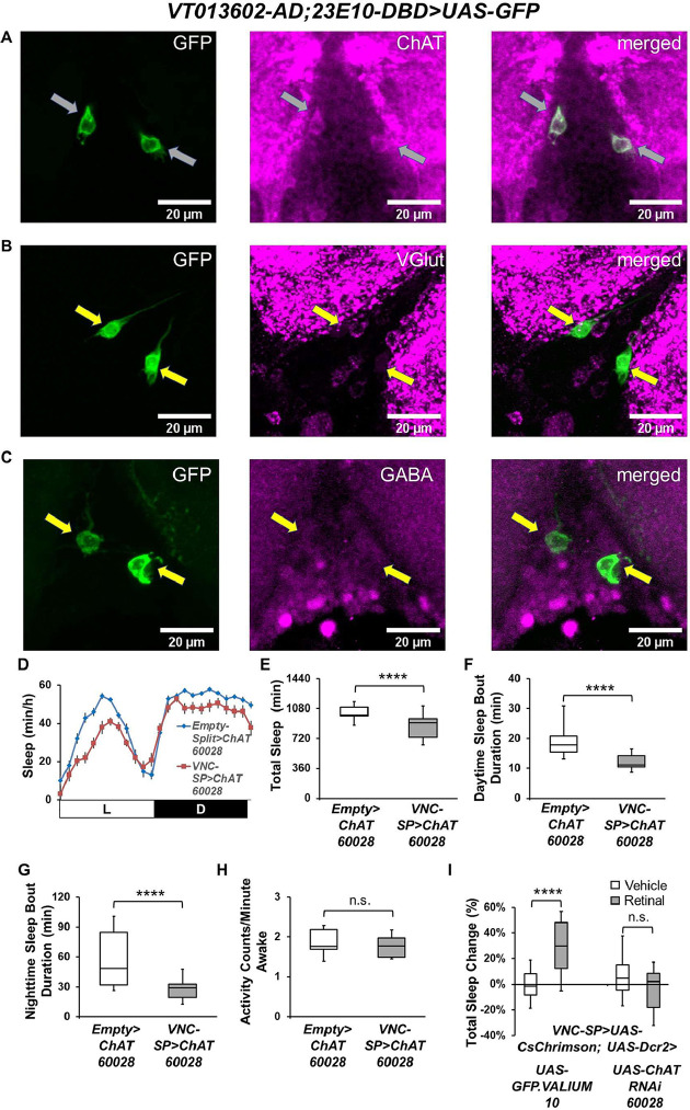
Fig 4. VNC-SP neurons are cholinergic.
(A–C) Representative confocal stacks focusing on the metathoracic ganglion of the VNC of female VT013602-AD; 23E10-DBD>UAS-GFP flies stained with antibodies to ChAT (A), VGlut (B), and GABA (C). Gray arrows in (A) indicate colocalization of GFP and ChAT staining in VNC-SP neurons. Yellow arrows in (B) and (C) indicate the localization of VNC-SP neurons. Green, anti-GFP; magenta, anti-ChAT (A), anti-VGlut (B), anti-GABA (C). (D) Sleep profile in minutes of sleep per hour for control (Empty-AD; 23E10-DBD>UAS-ChATRNAi 60028, blue line) and VNC-SP>ChATRNAi 60028 (VT013602-AD; 23E10-DBD>UAS-ChATRNAi 60028, red line) female flies. (E) Box plots of total sleep time (in minutes) for flies presented in (D). A two-tailed unpaired t test revealed that VNC-SP> ChATRNAi 60028 flies sleep significantly less than controls. ****P < 0.0001, n = 22–30 flies per genotype. (F) Box plots of daytime sleep bout duration (in minutes) for flies presented in (D). A two-tailed Mann–Whitney U test revealed that VNC-SP> ChATRNAi flies daytime sleep bout duration is significantly reduced compared with controls. ****P < 0.0001, n = 22–30 flies per genotype. (G) Box plots of nighttime sleep bout duration (in minutes) for flies presented in (D). A two-tailed Mann–Whitney U test revealed that VNC-SP> ChATRNAi flies nighttime sleep bout duration is significantly reduced compared with controls. ****P < 0.0001, n = 22–30 flies per genotype. (H) Box plots of locomotor activity counts per minute awake for flies presented in (D). A two-tailed Mann–Whitney U test revealed that there is no difference in waking activity between VNC-SP> ChATRNAi flies and controls, n.s. = not significant, n = 22–30 flies per genotype. (I) Box plots of total sleep change in % for vehicle-fed and retinal-fed VT013602-AD; 23E10-DBD>UAS-CsChrimson; UAS-GFP.VALIUM10 (control) and VT013602-AD; 23E10-DBD>UAS-CsChrimson; UAS-ChATRNAi 60028 flies stimulated by 627-nm LEDs. A two-way ANOVA followed by Sidak’s multiple comparisons shows that sleep is significantly increased in control flies and that expressing ChAT RNAi in VNC-SP neurons completely abolishes the sleep-promoting effect of VNC-SP neurons activated by 627-nm LEDs. ****P < 0.0001, n.s. = not significant, n = 31–36 flies per genotype and condition. The raw data underlying parts (E–I) can be found in S1 Data. VNC, ventral nerve cord; VNC-SP, VNC sleep-promoting.

