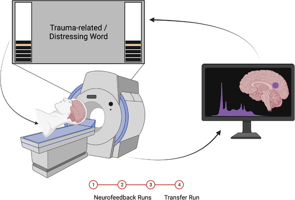
FIGURE 1.

Depiction of the rt‐fMRI‐NFB set‐up. While participants were inside the scanner, they were presented with a neurofeedback signal in the form of a virtual thermometer that increased/decreased in response to fluctuating activity within the neurofeedback target region (PCC). Participants completed three neurofeedback training runs, followed by a transfer run, in which they were not presented with the neurofeedback signal. Figure reproduced with permission from Nicholson et al. (2021).
