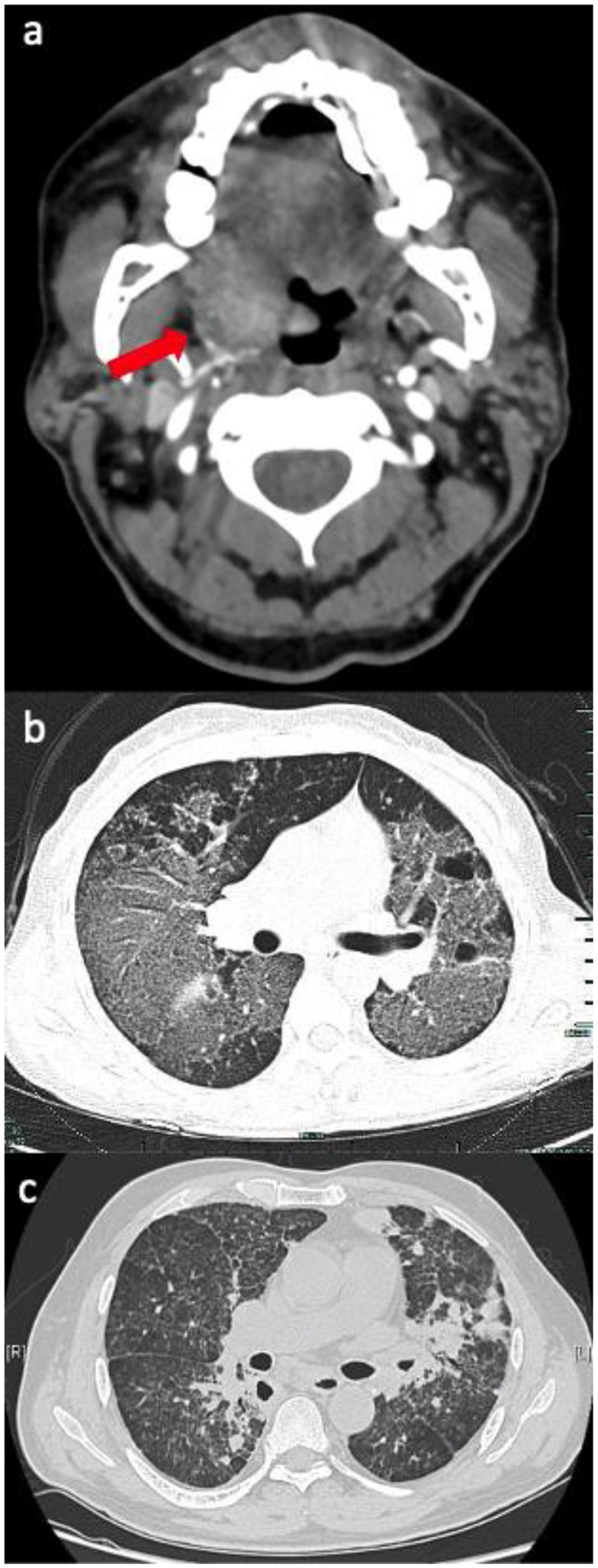
FIGURE 2.

Upper and lower respiratory tract manifestations of talaromycosis.
a) Computed tomography (CT) Angiogram of the neck demonstrating an ill-defined mass along the right lateral aspect of the hypopharynx involving the base of the tongue, right lingual tonsil, and right vallecula extending along the right palatine tonsil and into the pharyngeal space, in a 63 year-old man with HIV,1 b) Axial CT Chest demonstrating multiple disseminated ground glass opacities and bullae in a 34 year old immunocompromised female with a STAT3 mutation,2 c) Chest CT with interstitial infiltrates and nodules in a 57-year-old non-HIV-infected man with a history of prolonged steroid use.3
