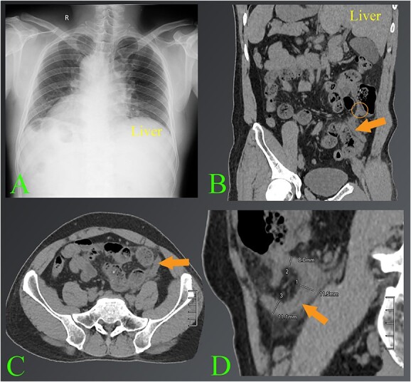
Figure 1.

The images of appendicitis in a patient with situs inversus. (A) Chest X-ray of the chest shows that the heart is on the right side. (B–D) The CT images show the left-sided acute appendicitis with surrounding fatty infiltration (arrows). The root of the appendix located high in the left lumbar position is also seen (circle). Note where the liver is located on the left side.
