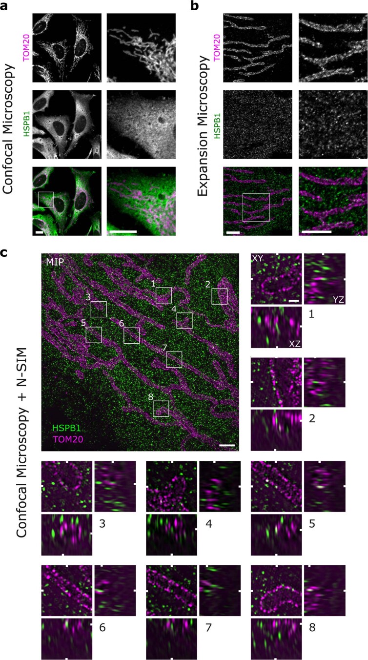
Extended Data Fig. 6. Mitochondrial-localized HSPB1 imaged using confocal microscopy, expansion microscopy, or a combination of expansion microscopy and structured illumination microscopy.
Non-heat shocked HeLa cells were labeled by immunofluorescence staining of endogenous HSPB1 and TOM20 as an outer mitochondrial membrane marker. All samples were first fixed and stained according to our standard protocol for confocal microscopy. For expansion microscopy, immunolabeled samples were linked to a 4x expandable hydrogel. a, Non-expanded samples were imaged with confocal laser scanning microscopy. Scale bar is 10 μm. b, Expansion microscopy samples imaged with confocal microscopy. Expansion-corrected scale bar is 2.5 μm c, Samples prepared as in (b) were imaged with structured illumination microscopy (SIM). The overview image is a maximum intensity projection (MIP) of the imaged volume and the zoomed boxes (1 µm x 1 µm) show examples of HSPB1 present inside mitochondria. White markers indicate the position of the orthogonal planes in the volumes (XY, XZ, and YZ) and were sliced at HSPB1 spots fully contained by the mitochondrial TOM20 boundaries in all dimensions. Expansion-corrected scale bar is 1.0 μm and 0.20 μm (zooms). Images are representative of one (c) and more than three replicates (a,b).

