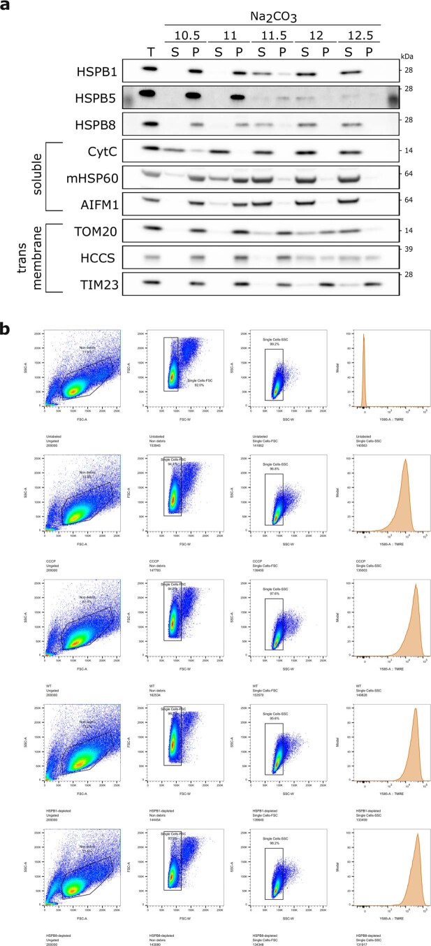
Extended Data Fig. 7. Small heat shock proteins detach from mitochondrial membranes around pH 11.5 and have no effect on mitochondrial membrane potential.
a, Membrane association of small heat shock proteins was verified with a sodium carbonate assay. This assay allows the separation of integral membrane proteins (retained in the pellet) from peripheral membrane proteins and soluble proteins (extracted into the supernatant). Mitochondria isolated from HeLa cells were resuspended in Na2CO3 set to the indicated pH. Samples were analyzed by SDS-PAGE followed by immunoblotting using anti-HSPB1, anti-HSPB5, anti-HSPB8, anti-CytC (IMS), anti-mtHSP60 (matrix), anti-AIFM1 (IMS), anti-TOM20 (OM), anti-HCCS (IM), and anti-TIM23 (IM) antibodies. T = total; P = pellet; S = supernatant. Unprocessed blots are available in source data. b, Flow cytometry gating strategy and TMRE membrane potential in control or sHSP-depleted HeLa cells. As a negative control, the membrane potential was dissipated by CCCP treatment. Related to Fig. 3d: ungated, non-debris, singlets, TMRE-intensity. FSC: forward scatter, SSC: side scatter, A: Area, H: height, W: width. Results are representative of one (b), in which more than 130,000 cells were counted per condition, or three (a) replicates. Unprocessed blots are available in source data.

