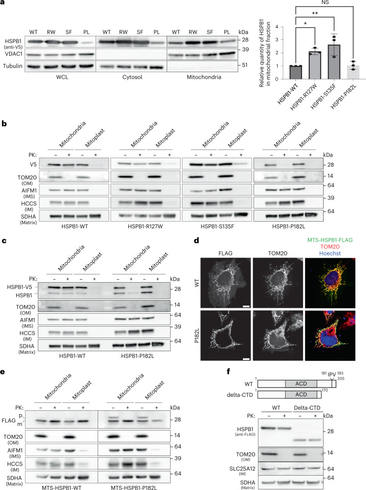
Fig. 6. CMT disease-causing mutations in HSPB1 disturb its mitochondrial function.
a, Mitochondria were isolated from HeLa cells overexpressing HSPB1 wild-type (WT), HSPB1 mutants R127W (RW), S135F (SF) or P182L (PL) and compared to the cytosol and the NP-40 soluble WCL. Note that the reduced expression of P182L in the WCL is due to reduced solubility in the NP-40-containing buffer. Samples were analysed by SDS–PAGE followed by immunoblotting using anti-V5 (HSPB1), anti-tubulin (cytosolic marker) and anti-VDAC1 (mitochondrial marker) antibodies. The graph represents the densitometric analysis of the mitochondrial fraction after correction for loading using VDAC1 (mean ± s.d.) (n = 3 biologically independent experiments). One-way ANOVA with Dunnett’s multiple comparison test was performed. *P < 0.05, **P < 0.01. NS, non-significant. b, Submitochondrial localization of HSPB1-V5 wild-type (WT) and HSPB1 mutants R127W, S135F and P182L was verified by subjecting intact mitochondria and mitoplasts (derived after osmotic swelling) to proteinase K (PK, 10 μg ml−1) treatment. Samples were analysed by SDS–PAGE followed by immunoblotting using anti-V5 (HSPB1), anti-TOM20 (OM), AIFM1 (IMS), HCCS (IM) and SDHA (matrix) antibodies. c, The same samples as in b were analysed with an anti-HSPB1 antibody to detect both endogenous and exogenous HSPB1. d, A fusion protein between HSPB1 and the MTS of COX8A was expressed in HeLa cells. Immunostaining with anti-FLAG (MTSCOX8a-HSPB1) and anti-TOM20 (mitochondrial marker). Scale bar, 10 μm. e, Submitochondrial localization of wild-type or P182L-mutant MTSCOX8a-HSPB1 was verified as in b. Samples were analysed by SDS–PAGE followed by immunoblotting using anti-FLAG (MTSCOX8a-HSPB1), anti-TOM20 (OM), anti-AIFM1 (IMS), anti-HCCS (IM) and anti-SDHA (matrix) antibodies. Precursor (p) and mature (m) proteins were separated by size as the N-terminal MTS was cleaved off after mitochondrial import. f, Proteinase K protection assay of mitochondria isolated from HeLa cells expressing full-length HSPB1 or C-terminally truncated HSPB1 (delta-CTD). Samples were analysed by SDS–PAGE followed by immunoblotting using anti-FLAG (HSPB1), anti-TOM20 (OM), SLC25A12 (IM) and SDHA (matrix) antibodies. The IPV motif is a conserved C-terminal motif, disrupted by the P182L mutation, and further described in ref. 81. Results are representative of three replicates (a–f). Source numerical data, including exact P values, and unprocessed blots are available in source data.

