Fig. 5.
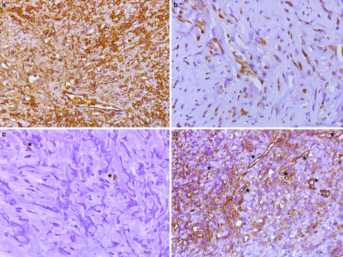
Immunohistochemistry: homogeneously positive reaction in the tumor cells for vimentin (a). Tumor cells showed a positive reaction for STAT6 (b). Immunohistochemistry for Ki-67 (Mib1), detected about 1% of all the tumor cells that were proliferating (c) with asterisks indicating two proliferating cells. Immunohistochemistry for CD34 (d) showed a positive reaction in the endothelial cells that constituted the dilated, branched, hyalinized staghorn-like (hemangiopericytoma-like) vasculature. Asterisks indicate a few exemplary vessels. Scale bars: 50 µm

