FIG. 6.
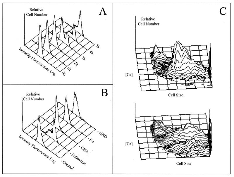
FCM analysis of poliovirus-infected HeLa cells. (A) Kinetics of intracellular free calcium during poliovirus infection. Poliovirus infection was performed at a multiplicity of infection of 50. After 1 h of adsorption at the indicated times postinfection, poliovirus-infected cells were detached from the plates, incubated with 6 μM fluo-3 AM, and analyzed in a FACScan flow cytometer. (B) Effects of guanidine, cycloheximide, and Ro 09-179 on [Ca2+]i during poliovirus infection. Guanidine (GND) (500 μM), Ro 09-179 (Ro) (1 μg/ml), or cycloheximide (CHX) (50 μM) was added to the infected cells after 1 h of poliovirus adsorption. The [Ca2+]i was monitored using FCM 4 h postinfection. Nontreated poliovirus-infected cells (Poliovirus) and nontreated uninfected cells (Control) are also shown. (C) Simultaneous FCM analysis of cytopathic effect and [Ca2+]i on poliovirus-infected cells. Noninfected (top) and poliovirus-infected (bottom) HeLa cells were analyzed 4 h postinfection for cytosolic free calcium (as described in panel A) and cell size (measured as forward light scatter). Adapted from reference 145 with permission of the publisher.

