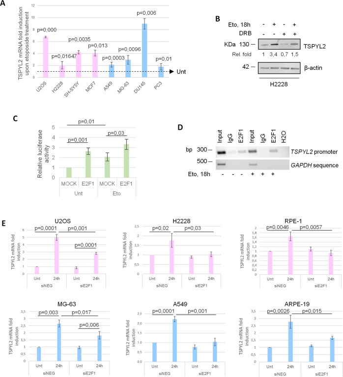
Fig. 2. E2F1 promotes TSPYL2 gene expression in response to DNA damage.
A RT-qPCR of TSPYL2 induction at 24 h after etoposide treatment in U2OS, H2228, SH-SY5Y, MCF7, A549, MG-63, DU145 and PC3 cell lines. P values were derived from Student’s t test between untreated and treated samples for each cell line. B Western blot analysis of TSPYL2 levels in H2228 cells treated with DRB (5,6-dichloro-1-beta-D-ribofuranosylbenzimidazole) and etoposide for 18 h in different combinations. C Luciferase assays of U2OS cells transfected with FLAG-E2F1 or empty vector (MOCK), before and after etoposide treatment for 6 h. P values were derived from Student’s t test between the indicated samples. D Chromatin immunoprecipitation (ChIP) assays of E2F1 on TSPYL2 promoter in untreated and etoposide-treated RPE-1 cells. Normal rabbit IgG were used as negative controls. Signals represent negative printing of ethidium bromide staining of PCR products. E RT-qPCR analysis of TSPYL2 mRNA levels before and after 24 h of etoposide treatment in control and E2F1 silenced U2OS, H2228, RPE-1, MG-63, A549 and ARPE-19 cell lines. P values were derived from Student’s t test between the indicated samples.

