
Figure 1. Bright field and immunocytochemical images in OrganoPlate® 2-lane 96 plate.
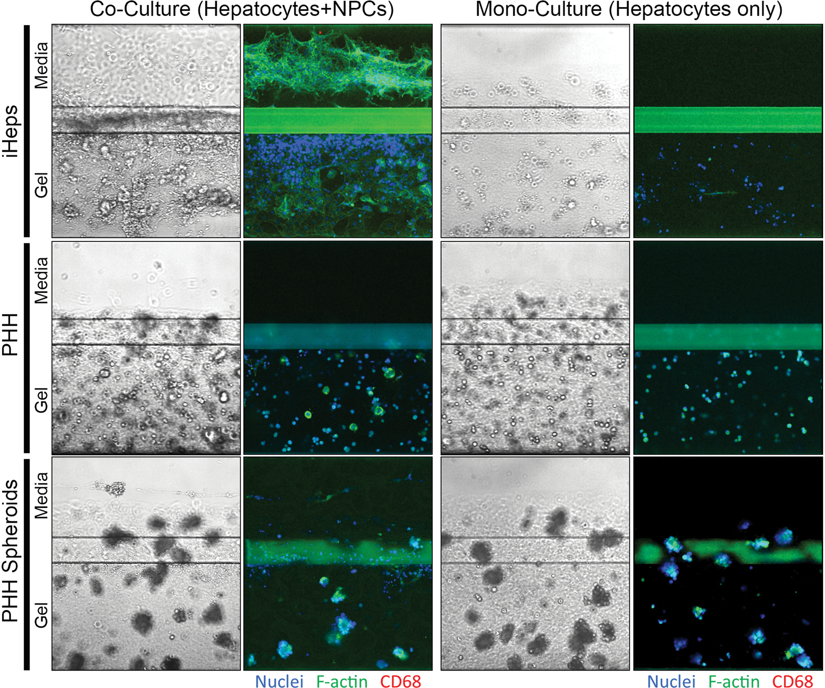
OrganoPlate® 2-lane 96 plates consist of the perfusion and organ lanes separated by a phase guide. Under all conditions, differentiated iHeps, primary human hepatocytes (PHH), or primary human hepatocyte spheroids were cultured in the gel lane with type 1 collagen. Under co-culture conditions, HMEC-1 endothelial cells and THP-1 derived macrophages were cultured in the perfusion lane. Images are shown for iHeps and primary human hepatocytes after 17 days of culture and for primary human hepatocyte spheroids after 14 days of culture. Some hepatocytes may appear to be on top of the Phaseguide or inside the perfusion channel; however, because the gel forms a meniscus they are still trapped in the gel. For immunocytochemistry, nuclei were stained with Hoechst-33342, cytoplasm was stained with F-actin, and THP-1 monocytes were stained with anti-CD68 antibodies.