. 2023 Jan 13;83(6):814–829. doi: 10.1158/0008-5472.CAN-22-1444

Figure 5.

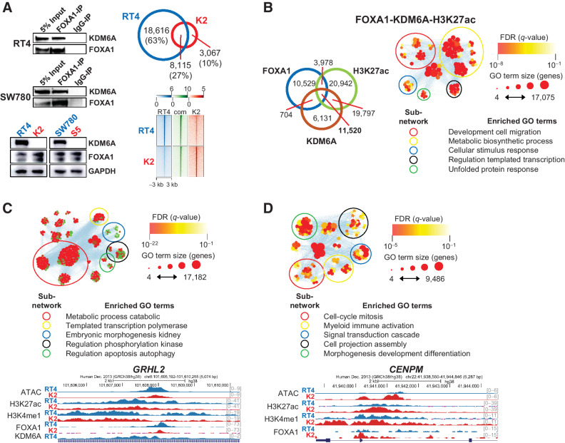
Figure 5. FOXA1 requires KDM6A in luminal subtype cells for the regulation of urothelial identity and homeostasis. A, Coimmunoprecipitation of KDM6A in using FOXA1 as a bait (top left). FOXA1 expression in parental (blue) and KDM6A knockout (red) cells (bottom left). Venn diagram showing shared and private FOXA1 peaks in RT4 parental and K2 knockout cells (top right). Heat map showing FOXA1 ChIP-seq signal at shared and private peaks (bottom right). B, Venn diagram showing KDM6A CUT&RUN, FOXA1 ChIP-seq, and H3K27ac ChIP-seq shared and private peaks in RT4 cells (left). Functional enrichment analysis of enhancer peaks bound by both FOXA1 and KDM6A (right). C, Functional enrichment analysis of FOXA1-bound genes that are upregulated in RT4 cells and genome browser view of the GRHL2 locus. D, Functional enrichment analysis of FOXA1-bound genes that are upregulated in K2 cells and genome browser view of the CENPM locus.

FOXA1 requires KDM6A in luminal subtype cells for the regulation of urothelial identity and homeostasis. A, Top left, coimmunoprecipitation of KDM6A in using FOXA1 as a bait. Bottom left, FOXA1 expression in parental (blue) and KDM6A knockout (red) cells. Top right, Venn diagram showing shared and private FOXA1 peaks in RT4 parental and K2 knockout cells. Bottom right, heat map showing FOXA1 ChIP-seq signal at shared and private peaks. B, Left, Venn diagram showing KDM6A CUT&RUN, FOXA1 ChIP-seq, and H3K27ac ChIP-seq shared and private peaks in RT4 cells. Right, functional enrichment analysis of enhancer peaks bound by both FOXA1 and KDM6A. C, Functional enrichment analysis of FOXA1-bound genes that are upregulated in RT4 cells and genome browser view of the GRHL2 locus. D, Functional enrichment analysis of FOXA1-bound genes that are upregulated in K2 cells and genome browser view of the CENPM locus.