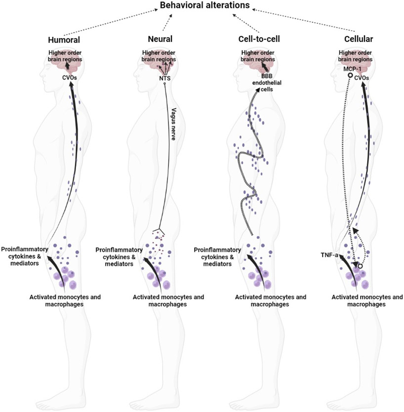
Figure 1.
Adapted from (Capuron and Miller, 2011). Peripheral-to-central immune signaling pathways. The humoral pathway involves peripherally released factors crossing the BBB (e.g., circumventricular organs; CVO) and directly influencing neuronal function and eliciting glial effects. The neuronal pathway involves the transduction of immune messages to a neural message in primary afferent neurons, which is then relayed to higher order brain regions via the nucleus tractus solitarius (NTS); vagal innervation and pro-inflammatory cytokine activity at peripheral nociceptors are examples of this. The cell-to-cell route relies on immune signals to roll along the blood vessel walls that engage with endothelial cells at the BBB, forming the interface between the blood and the brain. Endothelial cells become reactive and act as an immune signal translator to elicit central responses. Finally, the cellular route occurs when peripherally reactive pro-inflammatory cytokines, such as TNF-α, stimulate microglia which results in the release of factors such as monocyte chemoattractant type-1 (MCP-1) which recruits monocytes into the brain (D'Mello et al., 2009), and in specific brain nuclei to provide support for changes in neuronal function. Figure created in BioRender.com.

