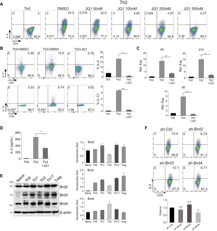
Figure EV1. Brd4 promotes Th2 cell differentiation.

- Flow cytometry analysis of mouse Th2 cells treated with JQ1 as indicated. Mouse Th2 cells were differentiated from mouse primary naïve CD4+ T cells for 6 days before analysis.
- Flow cytometric (left) and statistical analysis (right) of IL‐4 and IL‐13 in Th2 cells derived from mouse primary naïve CD4+ T cells treated with or without JQ1 (500 nM).
- qPCR analysis of Il4, Il5, and Il13 in mouse Th2 cells treated with or without JQ1.
- ELISA analysis of IL‐4 secretion into the supernatant of mouse Th2 cells treated with or without JQ1.
- Western blotting (left) and densitometry analysis (right) of Brd2, Brd3, and Brd4 in mouse naïve CD4+ T cells, Th0, Th1, Th2, Th17, and Treg cells.
- Flow cytometric (upper) and statistical analysis (lower) of IL‐4 in mouse Th2 cells infected with sh‐Ctrl, sh‐Brd2, sh‐Brd3, or sh‐Brd4 lentivirus.
Data information: Mouse naïve CD4+ T cells were cultured in Th2 polarization condition and treated with or without inhibitors on Day 0 and were differentiated for 6 days before analysis, unless otherwise specified. All data represent mean ± SD and average of three independent experiments. Data are analyzed by Paired t test. *P < 0.05; **P < 0.01; and ***P < 0.001.
Source data are available online for this figure.
