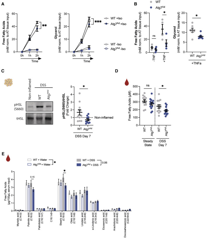
Figure 3. Autophagy loss reduces secretion of fatty acids from adipocytes.

- Ex vivo lipolysis measured by released free fatty acid (left, n = 4–5/group) and glycerol (right, n = 7–8/group) in culture supernatant of adipose tissue explants simulated with isoproterenol (10 μM) for 1–2 h.
- Ex vivo lipolysis measured by released free fatty acid (left, n = 4/group) and glycerol (right, n = 7/group) adipose tissue explants simulated with TNFα (100 ng/ml) for 24 h before replacing with fresh medium in the absence of TNFα for 3 h.
- Representative immunoblot for key lipolytic enzymes HSL, pHSL (Ser660) and quantification (n = 10–11/group).
- Serum levels of circulating FFAs measured in wild‐type and Atg7‐deficient mice (n = 13–14/group).
- Concentration of individual FFA species in serum in water‐treated and DSS‐treated mice as measured by FID‐GC (n = 12–14/group).
Data are represented as mean ± s.e.m. (A, B, E) Two‐Way ANOVA. (B, D) Unpaired Student's t‐test. (C) Mann–Whitney test. *P < 0.05, **P < 0.01, ***P < 0.001.
Source data are available online for this figure.
