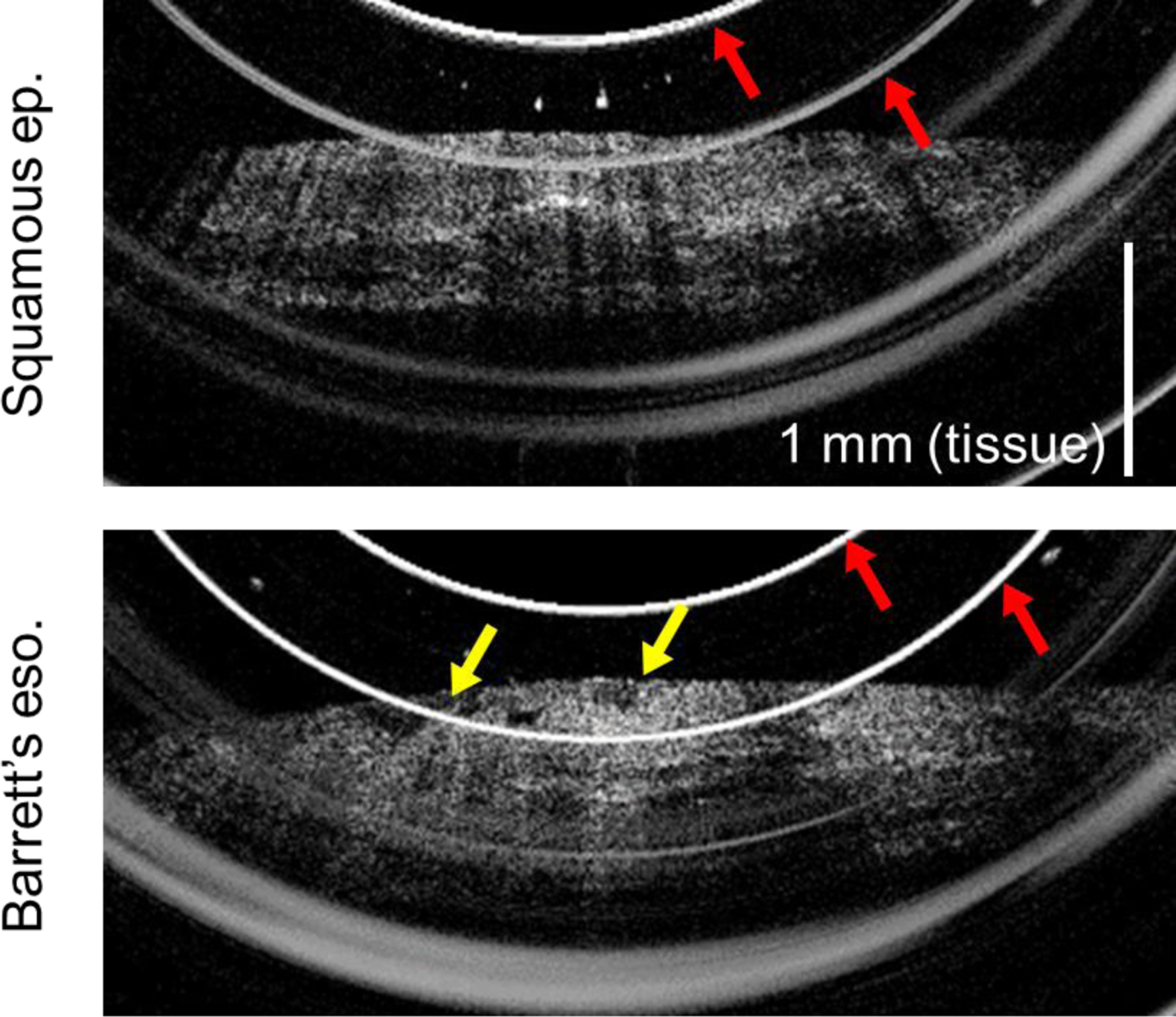
Figure 4:

OCT cross-sectional images of esophageal epithelium acquired by probe version 1. Red arrows indicate artifacts caused by internal probe reflections. Top: Squamous epithelium, showing smooth epithelial surface and consistent layered appearance. Bottom: Barrett’s esophagus, exhibiting an uneven nodular surface (yellow arrows).
