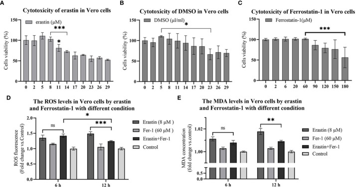
Figure 1.
16 hours cytotoxicity of Vero cells. ROS and lipid oxidation levels were used to evaluate the biological functions of erastin and Fer-1 in Vero cells. (A–C) Cytotoxicity of erastin, Ferrostatin-1 and DMSO in Vero cells at 16 hours. (D) The ROS levels of Vero cells treated with erastin and Fer-1 were detected at 6 h and 12 h. (E) Malondialdehyde levels in Vero cells treated with erastin and Fer-1 were detected at 6 and 12 hours. The data were performed from three independent experiments, with the untreated group as the control group. The differences were evaluated using ANOVA test. The error bar represents the standard deviation. ns, not significant, * P<0.05, ** P<0.01, *** P<0.001.

