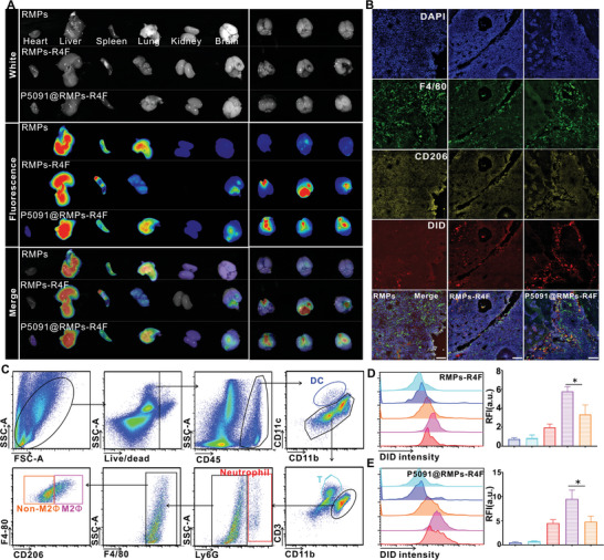
Figure 4.

P5091@RMPs‐R4F cross the blood brain barrier in vivo. A) A whole body imaging system was used to analyze the distribution of DiD dye‐stained RMPs, RMPs‐R4F, or P5091@RMPs‐R4F in different organs 24 h after i.v. injection. B) Immunofluorescence was performed to detect the CD206‐positive macrophages as an indication of the targeting ability of RMPs, RMPs‐R4F, and P5091@RMPs‐R4F injected into the tail vein. Images were obtained by confocal microscopy. Red‐DiD, Yellow‐CD206, Green‐F4/80, Blue‐DAPI. Scale bar: 100 µm. Data are presented as the mean ± SEM (n = 3). C) Gating strategy to distinguish different immune cell types. D,E) Fluorescence intensity of different immune cell types by flow cytometry. Statistical analysis was performed using one‐way ANOVA with Tukey's multiple comparison test (n = 3). Data are presented as the mean ± SEM. *p < 0.05, **p < 0.01, ***p < 0.001.
