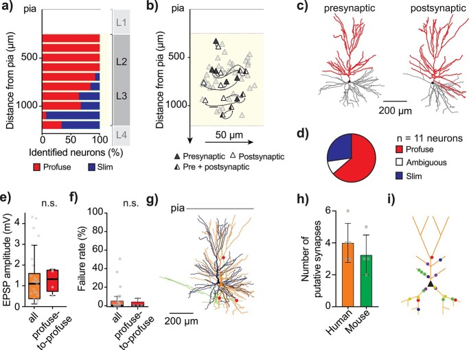
Fig. 3.
Morphological and anatomical analysis of local connections in L2/L3 of human MTG. a) Distribution of identified single neurons throughout L2/L3 of human MTG [total of 87 identified single neurons of which 60 were already identified for a previous study (Deitcher et al. 2017) and 27 were newly identified here], with profuse-tufted neurons (n = 58 neurons) in red and slim-tufted neurons (n = 29 neurons) in blue. b) Distribution within L2/L3 of recorded clusters where soma-pia distances were recorded through microdrive coordinates. X-axis distance between neurons of the same cluster is true to scale but X-axis distance between clusters is not and has been randomly chosen for clarity. c) Example morphological reconstruction (soma and apical dendrites in red, basal dendrites in gray) of 2 connected neurons (left: presynaptic, right: postsynaptic neuron). d) Pie graph showing number of identified neurons per cell-type (8 profuse-tufted neurons and 2 “ambiguous” type neurons, forming 4 profuse-to-profuse and 2 ambiguous-to-profuse type connections). e) Median EPSP amplitude of identified profuse-to-profuse connections relative to the full population of this study (excluding the identified profuse-profuse connections). f) Median fail rate of identified profuse-to-profuse connections relative to the full population of this study (excluding the identified profuse-profuse connections). g) Reconstructed connected pair of neurons from Fig. 1 (presynaptic neuron in black with axon in green, postsynaptic neuron in orange and putative synapse locations in red). Images of putative synapses for this connection in Fig. S6. h) Human presynaptic neurons formed on average 4.0 ± 1.2 putative synapses (n = 5 connections) onto their postsynaptic neuron comparable with mouse average number of putative synapses: 3.2 ± 1.1, (n = 4 connections). i) Schematic postsynaptic neuron depicting locations of all putative synapses from 5 distinct connections in L2/L3 human MTG. Putative synapses were located in basal dendrites and proximal apical dendrites (total mean distance from soma: 147 ± 54 μm) and were at similar dendritic distances to mouse (mouse total mean dendritic distance to soma: 141 ± 26 μm).

