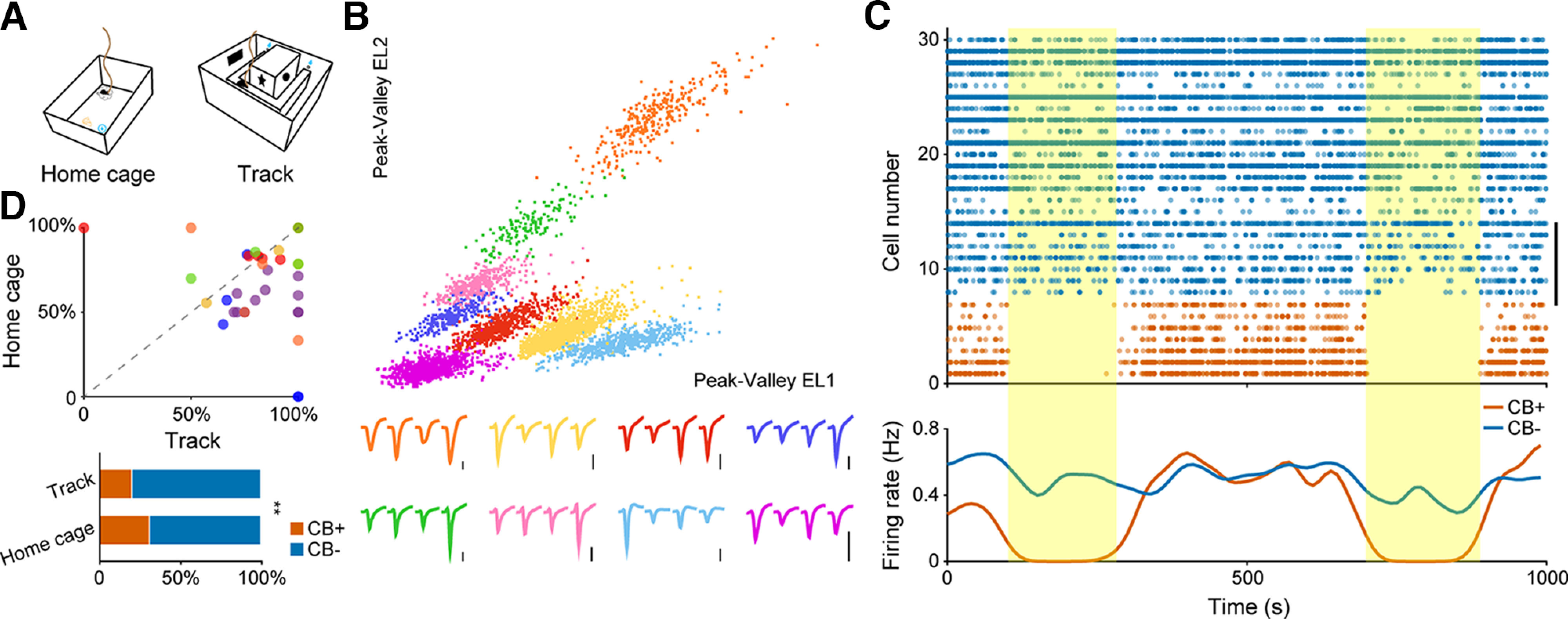
Figure 1.

In vivo identification and basic firing pattern of hippocampal pyramidal neuron subtypes. A, Two recording setups used in our experiment. Left, Home cage. Right, U-shape running track. B, Eight putative pyramidal neurons sorted from a single tetrode. Each color denotes one single unit. Corresponding tetrode waveforms were illustrated at the bottom. The orange cluster in the upper right corner represents one CB+ neuron. Scale bar: 0.2 mV. C, Neuronal firing sequence of 30 simultaneously recorded pyramidal neurons for a 1000-s recording period in home cage. Vertical bar on the right denotes eight neurons recorded from one single tetrode, as illustrated in B. Each dot represents one action potential. The firing of CB+ PNs were largely inhibited on optogenetic stimulation (yellow shaded areas). Bottom, Average firing rate of these neurons showing population responses of PNs to laser stimulation. Blue, CB− PNs; orange, CB+ PNs. D, Top, Ratio of CB− PNs recorded from each tetrode in running track as a function of that in home cage. Each dot represents result from one tetrode, while each color denotes individual animal (n = 6 mice). Bottom, Ratio of active CB+ and CB− PNs recorded from two recording setups. Note the significant decrease of the ratio of CB+ neurons recorded during running (**p = 6.6e-3, χ2 test).
